2.7.1.1: hexokinase
This is an abbreviated version!
For detailed information about hexokinase, go to the full flat file.
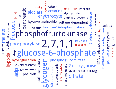
Word Map on EC 2.7.1.1
-
2.7.1.1
-
glucose-6-phosphate
-
phosphofructokinase
-
glycogen
-
fructose
-
citrate
-
adp
-
erythrocyte
-
malate
-
phosphorylase
-
pancreatic
-
hypoxia
-
islet
-
pentose
-
creatine
-
mellitus
-
2-deoxyglucose
-
aldolase
-
hyperglycemia
-
phosphoglucomutase
-
warburg
-
hypoxia-inducible
-
6-phosphogluconate
-
glut-1
-
voltage-dependent
-
vdacs
-
streptozotocin
-
positron
-
soleus
-
18f-fdg
-
lateralis
-
fructose-1,6-bisphosphatase
-
hypoglycemic
-
invertase
-
2-deoxy-d-glucose
-
fructokinase
-
gluconeogenic
-
insulin-stimulated
-
carboxykinase
-
hexoses
-
vastus
-
glycogenolysis
-
g6pase
-
glycosomes
-
antihyperglycemic
-
phosphoglucose
-
18f-fluorodeoxyglucose
-
fdg-pet
-
pharmacology
-
suvmax
-
analysis
-
glutaminolysis
-
2,6-bisphosphate
-
synthesis
-
diagnostics
-
industry
-
medicine
- 2.7.1.1
- glucose-6-phosphate
-
phosphofructokinase
- glycogen
- fructose
- citrate
- adp
- erythrocyte
- malate
- phosphorylase
- pancreatic
- hypoxia
- islet
- pentose
- creatine
- mellitus
- 2-deoxyglucose
- aldolase
- hyperglycemia
- phosphoglucomutase
-
warburg
-
hypoxia-inducible
- 6-phosphogluconate
-
glut-1
-
voltage-dependent
-
vdacs
- streptozotocin
-
positron
- soleus
-
18f-fdg
- lateralis
- fructose-1,6-bisphosphatase
-
hypoglycemic
- invertase
- 2-deoxy-d-glucose
- fructokinase
-
gluconeogenic
-
insulin-stimulated
-
carboxykinase
- hexoses
-
vastus
-
glycogenolysis
- g6pase
- glycosomes
-
antihyperglycemic
-
phosphoglucose
-
18f-fluorodeoxyglucose
-
fdg-pet
- pharmacology
-
suvmax
- analysis
-
glutaminolysis
-
2,6-bisphosphate
- synthesis
- diagnostics
- industry
- medicine
Reaction
Synonyms
6-phosphate glucose kinase, AtHXK1, ATP-D-hexose 6-phosphotransferase, ATP-D-hexose-6-phosphotransferase, ATP-dependent hexokinase, ATP: D-glucose 6-phosphotransferase, ATP: D-hexose 6-phosphotransferase, ATP:D-glucose 6-phosphotransferase, ATP:D-hexose 6-phosphotransferase, BmHk, brain form hexokinase, CzHXK1, FgHXK1, FgHXK2, GCK, GK, GKbeta, GlcK, glk, GlkB, glucokinase, glucokinase 1, glucokinase B, glucose ATP phosphotransferase, HaHXK1, hexokinase, hexokinase (phosphorylating), hexokinase 1, hexokinase 2, hexokinase 3, hexokinase 6, hexokinase A, hexokinase D, hexokinase Dor, hexokinase I, hexokinase II, hexokinase III, hexokinase IV, hexokinase PI, hexokinase PII, hexokinase type I, hexokinase type II, hexokinase type IV, hexokinase type IV glucokinase, hexokinase, tumor isozyme, hexokinase-1, hexokinase-10, hexokinase-2, hexokinase-4, hexokinase-5, hexokinase-6, hexokinase-7, hexokinase-8, hexokinase-9, hexokinase-II, hexokinase-like 1, hGK, hGK isoform 1, HK, HK II, HK-1, HK-I, HK-R, HK1, HK2, HK4, HKDC1, HKI, HKII, HKIII, HKL1, HPGLK1, HXK, HXK A, HXK II, Hxk1, HXK10, Hxk2, Hxk3, HXK4, HXK5, HXK6, HXK7, HXK8, HXK9, hxkC, hxkD, kinase, hexo- (phosphorylating), KlHxk1, LGK, LGK2, liver glucokinase, liver glucokinase isoform 2, MODY2 glucokinase, More, muscle form hexokinase, NfGlck, NtHXK1, OsHXK, OsHXK1, OsHXK10, OsHXK2, OsHXK3, OsHXK4, OsHXK5, OsHXK6, OsHXK7, OsHXK9, pHXK, plastid hexokinase, PpHXK1, PpHXK10, PpHXK11, PpHXK2, PpHXK3, PpHXK4, PpHXK5, PpHXK6, PpHXK7, PpHXK8, PpHXK9, Rag5p, ROK hexokinase, ScHK2, ScHXK2, SoHXK1, StHK, STK_23540, StoHK, TbHK1, TbHK2, TcHK, TthHK, type II hexokinase, VIT_18s0001g14230, xprF, Zm.5206, Zm.95484
ECTree

Advanced search results
Crystallization
Crystallization on EC 2.7.1.1 - hexokinase
Please wait a moment until all data is loaded. This message will disappear when all data is loaded.
 top
topCRYSTALLIZATION (Commentary) 

ORGANISM
UNIPROT
LITERATURE
homology modeling of structure reveals high structural homology with human glucokinase
crystal structure of hexokinase I from brain complexed with glucose and glucose 6-phosphate
-
first structures of a glucokinase-glucose complex without activator, of glucokinase-glucose-AMP-PNP and of glucokinase-glucose-AMP-PNP with a bound activator are reported. All structures are extremely similar, thus demonstrating that binding of GK activators does not result in conformational changes of the active protein but in stabilization of the active form of glucokinase
in complex with inhibitor 5-[[(2R,3S,4R,5R,6S)-5-[(3-bromophenyl)carbonylamino]-3,4,6-tris(oxidanyl)oxan-2-yl]methylsulfamoyl]-2-methyl-furan-3-carboxylic acid. Cocrystal structures reveal the flexibility of the hexokinase 2 protein and that the catalytic site can adopt an induced-fit conformation with inhibitors
molecular docking of 28-homobrassinolide. Homobrassinolide is able to bind to the drug-binding pocket of glucokinase. The glide energy is -7.1 kcal/mol
molecular docking of glucose and synthetic ligands (allosteric activators). Predicted position of glucose coincides with its position in the crystallographic complex. Glucose interacts with the residues of large (Ile225, Gly229, Cys230, Asn231, Glu256, Gln287, Glu290) and small (Ser151, Phe152, Pro153, Thr168, Lys169) domains and with the residues of connecting region Asn204, Asp205. Hydrogen bonds with Asp205, Glu256 and Glu290 stabilize this complex. The allosteric site formed by the small and large domains and two loops connecting them is located approximately 20 A away from the glucose binding site. There are two stable positions of glucokinase activators and approximately five sites of ligand binding with less favorable interaction energy. Predicted position of glucose coincides with its position in the crystallographic complex
multiple crystal forms of recombinant hexokinase I complexed with glucose/glucose 6-phosphate
-
purified recombinant hepatic wild-type and deletion mutant enzymes in complex with either D-glucose or the synthetic activator 2-amino-4-fluoro-5-(1-methyl-1H-imidazol-2-ylsulfanyl)-N-thiazol-2-yl-benzamide, hanging drop vapour diffusion method, 10 mg/ml protein in 20 mM Tris-HCl, pH 7.5, 50 mM NaCl, 5 mM Tris(2-carboxyethyl)phosphine hydrochloride, and 20 mM D-glucose, or 0.3 mM compound A, 0.0015-0.003 ml of the solution is mixed with an equal volume of precipitant solution containing 28-30% PEG 1500, 0.1 M HEPES-NaOH, pH 6.0, equilibration against 1 ml precipitant solution, 1 week, crystallization of the free hepatic enzyme by using precipitant solution containing 50 mM NaCl, 1.5-1.6 M ammonium sulfate, and 0.1 M bicine-NaOH, pH 8.7, 1 week, X-ray diffraction structure determination and analysis at 2.3 and 3.4 A resolution, respectively, molecular replacement
structure of HK2 in complex with glucose and glucose-6-phosphate. Crystals of DELTA16-hexokinase 2 are grown by sitting-drop vapor diffusion at 18°C
crystallization employing ammonium sulfate, diammonium phosphate or polyethylene glycol 6000 at pH values of 8.0-9.5 gives seven different crystal forms of KlHxk1. Crystallographic data to 1.66 A resolution are obtained using synchrotron radiation
-
in five crystal forms, a symmetrical ring-shaped homodimer is observed, corresponding to the physiological dimer existing in solution as shown by small-angle X-ray scattering. The dimer has a head-to-tail arrangement such that the small domain of one subunit interacts with the large domain of the other subunit. Dimer formation requires favorable interactions of the 15 N-terminal amino acids that are part of the large domain with amino acids of the small domain of the opposite subunit, respectively
sitting-drop vapour diffusion technique employing ammonium sulfate, diammonium phosphate or polyethylene glycol 6000 at pH values of 8.0-9.5 gives seven different crystal forms of KlHxk1. Crystallographic data to 1.66 A resolution are obtained using synchrotron radiation
-
sitting-drop vapour-diffusion method at 4°C. Structures of four different forms of OsHXK6 are presented in both open and closed state conformation
Crystals are grown in hanging drops, crystal structure of yeast hexokinase PI in complex with glucose and refined it at 2.95 A resolution
isoenzymes PI and PII, large crystals in the native and complexed form with glucose
-
hanging drop vapor diffusion method at 25°C. Crystal structures of StHK in four different forms: (i) apo-form, (ii) binary complex with glucose, (iii) binary complex with ADP, and (iv) quaternary complex with xylose, Mg2+, and ADP. Forms i and iii are in the open state, and forms ii and iv are in the closed state, indicating that sugar binding induces a large conformational change, whereas ADP binding does not
hanging drop vapor diffusion method. Crystal structures of StHK in four different forms: (i) apo-form, (ii) binary complex with glucose, (iii) binary complex with ADP, and (iv) quaternary complex with xylose, Mg2+, and ADP. Forms i and iii are in the open state, and forms ii and iv are in the closed state, indicating that sugar binding induces a large conformational change, whereas ADP binding does not
crystal structure of the enzyme is determined at a resolution of 2.02 A, with Rcryst and Rfree values of 18.1% and 22.6%, respectively
the crystal structure of the enzyme is determined at a resolution of 2.02 A