2.3.2.13: protein-glutamine gamma-glutamyltransferase
This is an abbreviated version!
For detailed information about protein-glutamine gamma-glutamyltransferase, go to the full flat file.
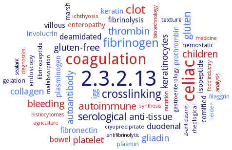
Word Map on EC 2.3.2.13
-
2.3.2.13
-
celiac
-
coagulation
-
clot
-
fibrinogen
-
crosslinking
-
gluten
-
serological
-
bleeding
-
thrombin
-
autoimmune
-
children
-
autoantibody
-
platelet
-
gluten-free
-
keratinocytes
-
anti-tissue
-
gliadin
-
collagen
-
duodenal
-
igg
-
bowel
-
deamidated
-
fibronectin
-
villous
-
keratin
-
fibrinolysis
-
plasminogen
-
isopeptide
-
involucrin
-
enteropathy
-
prothrombin
-
cornified
-
plasmin
-
hemostatic
-
endoscopy
-
marsh
-
gelation
-
texture
-
ichthyosis
-
fibrinopeptide
-
cryoprecipitate
-
gastroenterology
-
rheological
-
leiden
-
malabsorption
-
filaggrin
-
nutrition
-
analysis
-
agriculture
-
diagnostics
-
antifibrinolytic
-
medicine
-
synthesis
-
food industry
-
biotechnology
-
histiocytomas
-
sealant
-
2-antiplasmin
- 2.3.2.13
- celiac
- coagulation
- clot
- fibrinogen
-
crosslinking
- gluten
-
serological
- bleeding
- thrombin
- autoimmune
- children
- autoantibody
- platelet
-
gluten-free
- keratinocytes
-
anti-tissue
- gliadin
- collagen
- duodenal
- igg
- bowel
-
deamidated
- fibronectin
-
villous
- keratin
-
fibrinolysis
- plasminogen
-
isopeptide
- involucrin
- enteropathy
- prothrombin
-
cornified
- plasmin
-
hemostatic
-
endoscopy
-
marsh
-
gelation
-
texture
- ichthyosis
-
fibrinopeptide
-
cryoprecipitate
-
gastroenterology
-
rheological
- leiden
-
malabsorption
- filaggrin
- nutrition
- analysis
- agriculture
- diagnostics
-
antifibrinolytic
- medicine
- synthesis
- food industry
- biotechnology
- histiocytomas
-
sealant
-
2-antiplasmin
Reaction
Synonyms
AcTG-1, BmTGA, chloroplast transglutaminase, chlTGase, cold active transglutaminase, cold-active transglutaminase, EPB42, factor XIII, factor XIIIa, fibrin stabilizing factor, fibrinoligase, Galphah, glutamine:amine gamma-glutamyl-transferase, glutaminylpeptide gamma-glutamyltransferase, glutamyltransferase, glutaminylpeptide gamma-, gpTGase 2, gTG2, hfXIIIa, hTG2, hTGase 1, hTGase 2, hTGase 3, hTGase 6, KALB, KalbTG, KALB_7456, Laki-Lorand factor, mammalian transglutaminase, microbial transglutaminase, microbial transglutaminases, MsTGase, MTG, MTG-TX, MTGase, mTGase 2, OlTGT, plastidial transglutaminase, polyamine transglutaminase, protein 4.2, protein-glutamine gamma-glutamyltransferase, R-glutaminyl-peptide:amine gamma-glutamyl transferase, R-glutaminylpeptide-amine gamma-glutamyltransferase, SCTG, SMTG, STG I, t-TG, TG-2, TG1, TG2, TG3, TG4, TG5, TG6, TG7, tGA, TGase, TGase 1, TGase 2, Tgase 3, TGase 6, Tgase II, TGase-2, TGase2, TGB, TGK1, TGK2, TGL, TGM1, TGM2, TGM3, TGM4, TGM5, TGM6, TGM7, TGZ, tgz15, TGZo, tissue transglutaminase, tissue transglutaminase 2, tissue-TG, transglutaminase, transglutaminase 1, transglutaminase 2, transglutaminase 3, transglutaminase 6, transglutaminase C, transglutaminase factor XIII, transglutaminase type II, transglutaminase-2, transglutaminase-like protein, transglutaminase2, tTG, tTG-2, type 2 transglutaminase, type I transglutaminase, type-2 transglutaminase
ECTree

Advanced search results
Crystallization
Crystallization on EC 2.3.2.13 - protein-glutamine gamma-glutamyltransferase
Please wait a moment until all data is loaded. This message will disappear when all data is loaded.
 top
topCRYSTALLIZATION (Commentary) 

ORGANISM
UNIPROT
LITERATURE
sitting drop method, X-ray diffraction data are collected using synchrotron radiation to 2.1 A resolution. Crystals of Tgl belong to the tetragonal space group P4(1,3) and contain two molecules per asymmetric unit
-
homology modeling of enzyme photolabeled by 3-(3-methyl-3H-diaziren-3-yl)-N-[4-[(1E)-3-oxo-3-(pyridin-3-yl)prop-1-en-1-yl]phenyl]propanamide. In the closed conformation of enzyme, C230 is more solvent-exposed than in the open conformation. Docking experiments suggest binding modes that block access to the tunnel leading to the active site
-
crystallization of the enzyme inhibited with Ac-P(6-diazo-5-oxo-L-norleucine)LPF-NH2, hanging-drop method at 25°C, 2 A resolution
hanging drop vapor diffusion method, using 20 mM MES at pH 6.8, 200 mM sodium chloride, 20 mM MgCl2, 6% (w/v) PEG 3350, 5 mM dithiothreitol, and 24% (v/v) glycerol
-
hanging drop vapour diffusion, 15 mg/ml transglutaminase 3, 0.1 mM beta-octylglucoside in 0.1 ml of enzyme in 20 mM Tris-HCl, pH 8.0, 1 mM EDTA, 125 mM NaCl and 3 mM CaCl2, well solution containing 4-12% polyethylene glycol 6K, 100 mM bicine, pH 9.0, and 1% dioxane at 21°C, X-ray structure of zymogen and activated transglutaminase 3 at 2.2 and 2.1 A resolution
human TG2 has been crystallized in its GDP- and GTP-bound form (PDB IDs 1KV3, 4PYG), in complex with ATP (PDB ID 3LY6), and with alternative inhibitors bound to its transamidase site within the alpha/beta-catalytic domain (PDB IDs 2Q3Z, 3S3J, 3S3P, 3S3S), structure modeling, analysis, overview
purified recombinant His-tagged enzyme, hanging drop vapor diffusion method, mixing of 0.001 ml of 10 mg/ml protein in 20 mM Tris, pH 8.0, and 150 mM NaCl with 0.001 ml of reservoir solution containing 20 mM MES, pH 6.2, 150 mM sodium chloride, 5mM MgCl2, 4% PEG 3350, and 5 mM DTT, and 20% glycerol, equilibration against 0.4 ml of the reservoir solution, 20°C, X-ray diffraction structrue determination and analysis at 3.2 A resolution
purified recombinant soluble KalbTG is crystallized at 22°C using the sitting drop vapor diffusion method by 1:1 mixing of 200 nl of 8 mg/ml protein in PBS with an unbuffered reservoir consisting of 0.2 M ammonium tartrate, and 20% PEG 3350, X-ray diffraction structure determination and analysis at 1.9 A resolution, structure modeling, the first 19 N-terminal amino acids (MGGGSTTAQAAAVAAPTPR) and the C-terminal artificial GGGSHis8 tag are disordered in the structure
hanging drop vapor diffusion method, using 0.2 M sodium chloride, 0.1 M HEPES, pH 7.2, 20% (w/v) polyethylene glycol 4000
docking model of MTG binding to substrate N-carbobenzoxy-L-glutaminyl-glycine. Substrate is stretched along the MTG active site cleft with hydrophobic and/or aromatic residues interacting directly with the substrate. An oxyanion binding site for TGase activity may be constructed from the amide groups of Cys64 and/or Val65
hanging drop vapor diffusion method, using 30% (w/v) PEG 8000, 50 mM NaCl, 1 mM EDTA, 1 mM 2-mercaptoethanol, 0.01% (w/v) NaN3, 100 mM cacodylic acid (pH 5.0), and 2% (v/v) glycerol
-