3.5.4.1: cytosine deaminase
This is an abbreviated version!
For detailed information about cytosine deaminase, go to the full flat file.
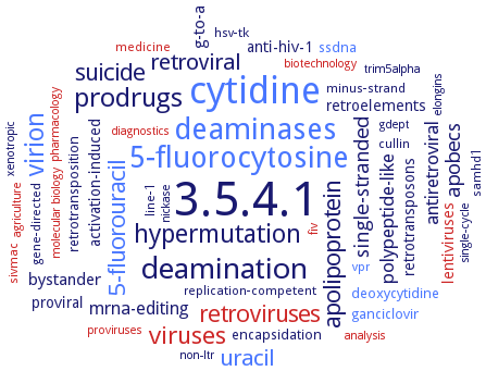
Word Map on EC 3.5.4.1
-
3.5.4.1
-
cytidine
-
deaminases
-
5-fluorocytosine
-
deamination
-
prodrugs
-
virion
-
hypermutation
-
viruses
-
5-fluorouracil
-
apolipoprotein
-
retroviruses
-
suicide
-
uracil
-
retroviral
-
apobecs
-
single-stranded
-
antiretroviral
-
polypeptide-like
-
mrna-editing
-
g-to-a
-
bystander
-
activation-induced
-
retrotransposons
-
anti-hiv-1
-
proviral
-
retroelements
-
lentiviruses
-
encapsidation
-
ganciclovir
-
deoxycytidine
-
retrotransposition
-
ssdna
-
fiv
-
samhd1
-
cullin
-
minus-strand
-
replication-competent
-
gene-directed
-
sivmac
-
medicine
-
line-1
-
hsv-tk
-
vpr
-
gdept
-
non-ltr
-
molecular biology
-
xenotropic
-
analysis
-
pharmacology
-
biotechnology
-
trim5alpha
-
diagnostics
-
proviruses
-
nickase
-
agriculture
-
single-cycle
-
elongins
- 3.5.4.1
- cytidine
- deaminases
- 5-fluorocytosine
-
deamination
-
prodrugs
- virion
-
hypermutation
- viruses
- 5-fluorouracil
-
apolipoprotein
- retroviruses
-
suicide
- uracil
-
retroviral
-
apobecs
-
single-stranded
-
antiretroviral
-
polypeptide-like
-
mrna-editing
-
g-to-a
-
bystander
-
activation-induced
-
retrotransposons
-
anti-hiv-1
-
proviral
-
retroelements
- lentiviruses
-
encapsidation
- ganciclovir
- deoxycytidine
-
retrotransposition
- ssdna
- fiv
- samhd1
-
cullin
-
minus-strand
-
replication-competent
-
gene-directed
- sivmac
- medicine
-
line-1
-
hsv-tk
- vpr
-
gdept
-
non-ltr
- molecular biology
-
xenotropic
- analysis
- pharmacology
- biotechnology
- trim5alpha
- diagnostics
- proviruses
-
nickase
- agriculture
-
single-cycle
-
elongins
Reaction
Synonyms
A3DE, APOBEC1, APOBEC3, APOBEC3G, CD, CDA, CDase, codA, CodA protein, Cytosine aminohydrolase, cytosine deaminase, cytosine deaminase I, cytosine deaminase II, cytosine deaminase P, cytosine deaminase S, cytosine deaminase Y, Fca1p, FCY1, isocytosine deaminase, yCD, Zn2+CDase
ECTree

Advanced search results
Crystallization
Crystallization on EC 3.5.4.1 - cytosine deaminase
Please wait a moment until all data is loaded. This message will disappear when all data is loaded.
 top
topCRYSTALLIZATION (Commentary) 

ORGANISM
UNIPROT
LITERATURE
10 mg/ml purified recombinant His-tagged enzyme, vapour phase equilibration against 11-14% PEG 8000, 0.1 M HEPES, pH 7.3-7.7, 0.2 M MgCl2, in hanging drop geometry, flash-cooling in a buffer containing 30% v/v glycerol for cryoprotection, also crystallization of a seleno-methionine derivatized enzyme, X-ray diffraction structure determination and analysis at 1.5 A resolution
the structure of Zn-CDA is determined to a resolution of 1.7 A with phosphonocytosine a potent mimic of the putative tetrahedral intermediate bound in the active site
-
the wild type enzyme in complex with the mechanism-based inhibitors 4-(R)-hydroxyl-3,4-dihydropyrimidine or 5-fluoro-4-(S)-hydroxyl-3,4-dihydropyrimidine and the mutant enzyme V152A/F316C/D317G are crystallized by the sitting drop vapor diffusion method, using 10-15% (w/v) PEG 6K, 200 mM MgCl2 and 100 mM HEPES (pH 7-8)
-
wild-type enzyme and mutants D314G, D314A, D314S, free and in complex with mechanism-based inhibitor 5-fluoro-4-(S)-hydroxyl-3,4-dihydropyrimidine
-
crystallization strategy named microseed matrix screening, differential chelation pattern of cations by acidic surfaces of proteins within crystal lattice as a critical parameter of crystal nucleation and growth
-
molecular dynamics simulation of free enzyme and in complex with cytosine, uracil and reaction intermediates
-
PDB ID 1UAQ, structure analysis and comparison to the quantum mechanical/molecular mechanical molecular dynamics simulation model, overview
purified recombinant wild-type and selenomethionine-labeled enzymes in 10% 2-propanol, 20% PEG 4000, 0.1 M Na HEPES, pH 7.5, at 4°C and at 22°C by hanging drop vapour diffusion method, 0.002 ml of both reservoir and protein solution are mixed in presence of 2-hydroxypyrimidine, 3-5 days to 1-2 weeks at 22°C, micro-seeding, X-ray diffraction structure determination and analysis at beyond 1.5 A resolution
-
recombinant A23L/D92E/V108I/I140L mutant bound to the transition state analogue, hanging drop vapour diffusion method, 2 days, crystals are soaked for 20 min in a mother liquor solution containing 2-hydroxypyrimidine concentrated 1.2:1 relative to protein, after soaking, the crystals are immediately transferred briefly to a cryo-buffer containing the 2-hydroxypyrimidine mother liquor plus 25% DMSO, X-ray diffraction structure determination and analysis at 2.3 A resolution, modelling and molecular replacement
the crystal structure of cytosine deaminase combined with the substrate uracil, PDB ID code: 1P6O, optimization in the water solvent at the ONIOM molecular dynamics study, overview