5.1.3.2: UDP-glucose 4-epimerase
This is an abbreviated version!
For detailed information about UDP-glucose 4-epimerase, go to the full flat file.

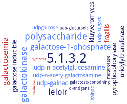
Word Map on EC 5.1.3.2 
-
5.1.3.2
-
polysaccharide
-
galactokinase
-
leloir
-
galactosemia
-
galactose-1-phosphate
-
udp-galnac
-
udp-n-acetylglucosamine
-
galactose-inducible
-
kluyveromyces
-
pyrophosphorylase
-
fragilis
-
uridylyltransferase
-
udp-n-acetylgalactosamine
-
udpglucose
-
udp-sugars
-
galnac
-
mutarotase
-
galactose-containing
-
o-antigens
-
udp-glucuronic
-
medicine
-
synthesis
- 5.1.3.2
- polysaccharide
- galactokinase
-
leloir
- galactosemia
- galactose-1-phosphate
- udp-galnac
- udp-n-acetylglucosamine
-
galactose-inducible
-
kluyveromyces
-
pyrophosphorylase
- fragilis
-
uridylyltransferase
- udp-n-acetylgalactosamine
- udpglucose
- udp-sugars
- galnac
- mutarotase
-
galactose-containing
-
o-antigens
-
udp-glucuronic
- medicine
- synthesis
Reaction
Synonyms
4-Epimerase, 6xHis-rGalE, ABD1_580, An14g03820, AtUGE1, AtUGE2, AtUGE3, AtUGE4, AtUGE5, CaGAL10, CapD, epimerase Ab-WbjB, Epimerase, uridine diphosphoglucose, fnlA, GAL10, Gal10p, Galactowaldenase, GalE, galE-1, galE-2, galE1, GalESp1, GalESp2, GNE, GNE2, H3634, HvUGE1, HvUGE2, HvUGE3, MdUGE1, More, OsUGE-1, OsUGE1, PsUGE1, Rdh1, rGalE, Rv3634c, TbGalE, TM0509, TMGalE, UDP-D-galactose 4-epimerase, UDP-D-glucose/UDP-D-galactose 4-epimerase, UDP-Gal 4-epimerase, UDP-Gal/Glc 4-epimerase, UDP-galactose 4'-epimerase, UDP-galactose 4-epimerase, UDP-galactose-4'-epimerase, UDP-galactose-4-epimerase, UDP-Glc 4-epimerase, UDP-Glc(NAc) 4-epimerase, UDP-GlcNAc 4-epimerase, UDP-GlcNAc/Glc 4-epimerase, UDP-glucose 4'-epimerase, UDP-glucose 4-epimerase, UDP-glucose 4-epimerase 1, UDP-glucose 4-epimerase 4, UDP-glucose epimerase, UDP-glucose-4-epimerase, UDP-glucose/-galactose 4-epimerase, UDP-hexose 4-epimerase, UDP-N-acetyl-glucosamine 4,6-dehydratase, UDP-sugar 4-epimerase, UDP-Xyl 4-epimerase, UDP-xylose 4-epimerase, UDPG-4-epimerase, UDPgalactose 4-epimerase, UGE, UGE-1, UGE1, UGE1:GUS, Uge1p, UGE2, UGE3, UGE3:GUS, UGE4, UGE5, UgeA, Uridine diphosphate galactose 4-epimerase, Uridine diphosphate glucose 4-epimerase, uridine diphosphate-galactose-4'-epimerase, Uridine diphospho-galactose-4-epimerase, Uridine diphosphogalactose-4-epimerase, Uridine diphosphoglucose 4-epimerase, Uridine diphosphoglucose epimerase, uridine-diphospho-glucose 4-epimerase, WbjB


 results (
 results (
 top
top


