1.14.11.29: hypoxia-inducible factor-proline dioxygenase
This is an abbreviated version!
For detailed information about hypoxia-inducible factor-proline dioxygenase, go to the full flat file.
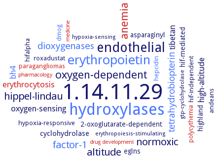
Word Map on EC 1.14.11.29
-
1.14.11.29
-
hydroxylases
-
endothelial
-
erythropoietin
-
anemia
-
oxygen-dependent
-
hippel-lindau
-
factor-1
-
normoxic
-
altitude
-
dioxygenases
-
tetrahydrobiopterin
-
oxygen-sensing
-
erythrocytosis
-
high-altitude
-
bh4
-
tibetan
-
cyclohydrolase
-
hif-mediated
-
eglns
-
asparaginyl
-
2-oxoglutarate-dependent
-
highland
-
hifalpha
-
paragangliomas
-
hepcidin
-
hypoxia-responsive
-
hif-independent
-
andeans
-
polycythemia
-
roxadustat
-
dmog
-
hypoxia-sensing
-
gtp-cyclohydrolase
-
erythropoiesis-stimulating
-
drug development
-
pharmacology
-
medicine
- 1.14.11.29
- hydroxylases
- endothelial
- erythropoietin
- anemia
-
oxygen-dependent
-
hippel-lindau
- factor-1
-
normoxic
-
altitude
- dioxygenases
- tetrahydrobiopterin
-
oxygen-sensing
- erythrocytosis
-
high-altitude
- bh4
-
tibetan
-
cyclohydrolase
-
hif-mediated
-
eglns
-
asparaginyl
-
2-oxoglutarate-dependent
-
highland
-
hifalpha
- paragangliomas
- hepcidin
-
hypoxia-responsive
-
hif-independent
-
andeans
- polycythemia
-
roxadustat
- dmog
-
hypoxia-sensing
-
gtp-cyclohydrolase
-
erythropoiesis-stimulating
- drug development
- pharmacology
- medicine
Reaction
Synonyms
Egl nine homolog 1, EGLN, EGLN1, Egln2, EGLN3, factor inhibiting HIF, FIH, HIF hydroxylase, HIF prolyl, HIF prolyl 4-hydroxylase, HIF prolyl hydroxylase, HIF-1alpha PHD3, HIF-1alpha prolyl hydroxylase 3, HIF-alpha prolyl-hydroxylase, HIF-P4H-1, HIF-P4H-2, HIF-P4H-3, HIF-PH, Hif-prolyl hydroxylase, HIF-prolyl hydroxylase domain 2, HIF-prolyl hydroxylase-2, HPH-1, HPH-2, HPH-3, hydroxylase domain enzyme, hypoxia-inducible factor prolyl hydroxylase 2, hypoxia-inducible factor prolyl hydroxylase domain 2, P4H-TM, PHD, PHD1, PHD2, PHD3, proline hydroxylase domain 2, prolyl 4-hydroxylase, prolyl hydroxylase, prolyl hydroxylase domain, prolyl hydroxylase domain protein, prolyl hydroxylase domain protein 2, prolyl hydroxylase-2, prolyl-4-hydroxylase 2, prolyl-4-hydroxylase domain 2, transmembrane prolyl 4-hydroxylase
ECTree

Advanced search results
General Information
General Information on EC 1.14.11.29 - hypoxia-inducible factor-proline dioxygenase
Please wait a moment until all data is loaded. This message will disappear when all data is loaded.
 top
topGENERAL INFORMATION 

ORGANISM
UNIPROT
COMMENTARY 
LITERATURE
drug target
prolyl 4-hydroxylase domain protein 3 (PHD3) overexpression has therapeutic potential in treatment of obstructive sleep apnea and intermittent hypoxia induced cardiovascular fibrosis
evolution
malfunction
metabolism
physiological function
additional information
evolution
the enzyme belongs to the of the 2-oxoglutarate- and iron-dependent dioxygenase family of enzymes
malfunction

-
silencing FIH, EC 1.14.11.30, under conditions where prolyl hydroxylase, is inhibited results in increased HIF-1alpha transcriptional activity, but paradoxically decreases HIF-1alpha stability
malfunction
-
hypoxia-inducible factor prolyl hydroxylase 2 knockdown leads to a less sustained activation of epidermal growth factor receptor and its downstream signaling pathways
malfunction
-
inactivation of Phd2 in endothelial cells specifically results in severe pulmonary hypertension but not polycythemia and is associated with abnormal muscularization of peripheral pulmonary arteries and right ventricular hypertrophy
malfunction
isoform PHD3 silencing leads to downregulation of most glycolytic enzymes from glucose transport to lactate production supported by the reduction in extracellular acidification and lactate production and increase in cellular oxygen consumption rate
malfunction
macrophages deficient in PHD3 have decreased levels of stress-induced apoptosis. The antiapoptotic effects of PHD3 knockout are independent of alterations in HIF and instead, appear to occur via reduced expression of Angptl2, an extracellular protein that is structurally similar to angiopoietins. Hypoxia-dependent PHD3 inhibition in macrophages promotes cell survival through the activation of HIF-dependent adaptive pathways and HIF-independent antiapoptotic pathways, including decreased expression of Angptl2, impact of altered PHD3 expression/activity on macrophage function, schematic overview
malfunction
PHD2 knockdown causes a marked reduction of erythropoietin (EPO) production. HIF seems not to be involved in this process
malfunction
the gene expression of p50 is reduced in the PHD2 knockdown cells, while the protein amount and the subcellular distribution of p65 are not changed in the PHD2 knockdown cells. The transactivation activity of NFkappaB is consequently decreased in the PHD2 knockdown cells. The expression levels of HIF target genes GLUT1 and VEGF-A is significantly upregulated in the PHD2 knockdown cells. The gene expression of BNIP3 is decreased. The influence of PHD2 knockdown on the gene expression of HIF-1alpha, HIF-2alpha, and p50 is not caused by off-target effect
metabolism
-
HIF protein stability is controlled by the oxygen sensing prolyl hydroxylase domain enzymes. Hypoxia-induced HIF signalling, mathematical modelling of the pathway, temporal dynamics of the HIF response to hypoxia, and molecular interaction map for the HIF network, overview. The hypoxia inducible factor is switched on and promotes adaptation to hypoxia by upregulatinggenes involved in angiogenesis, erythropoiesis and glycolysis
metabolism
-
hypoxia and oxidant stress can interact functionally as distinct regulators of HIF transcriptional output involving the enzyme. Oxidant stress activates hypoxia pathways through the inactivation of the oxygen-sensing hypoxia-inducible factor prolyl and asparaginyl hydroxylases
metabolism
-
optimal HIF-1alpha transcriptional activity requires sequential inhibition of both prolyl- and asparaginyl-hydroxylases
metabolism
-
isoforms PHD2 and 3 inhibit IkappaB kinase and decrease the activity of the nuclear factor-kappaB pathway. Isoform PHD2 also acts as a negative regulator of nuclear factor-kappaB during arteriogenesis
metabolism
-
isoforms PHD2 and 3 inhibit IkappaB kinase and decrease the activityof the nuclear factor-kappaB pathway. Isoform PHD2 also acts as a negative regulator of nuclear factor-kappaB during arteriogenesis
metabolism
PDH2 regulates hypoxia-inducible factor (HIF)-independent pathways, as well as the degradation pathway of HIFalpha. HIF-2alpha does not play a major role in the regulation of erythropoietin (EPO) expression by PHD2
metabolism
PHD isoforms have a differential contribution in controlling hypoxia-inducible factor (HIF)-alpha degradation and activity
metabolism
PHD isoforms have a differential contribution in controlling hypoxia-inducible factor (HIF)-alpha degradation and activity
metabolism
PHD isoforms have a differential contribution in controlling hypoxia-inducible factor (HIF)-alpha degradation and activity. Hydroxylases are key oxygen sensors expressed in all cells that regulate the adaptive response to hypoxia and promote a return to oxygen homeostasis. Complex crosstalk exists between inflammatory and hypoxic signaling pathways
physiological function
-
HIF (hypoxia-inducible factor) is a transcription factor that plays a pivotal role in cellular adaptation to changes in oxygen availability. In the presence of oxygen, HIF is targeted for destruction by an E3 ubiquitin ligase containing the von Hippel-Lindau tumor suppressor protein (pVHL). Human pVHL binds to a short HIF-derived peptide when a conserved proline residue at the core of this peptide is hydroxylated. This protein modifiation may play a key role in mammalian oxygen sensing
physiological function
-
hypoxia-inducible factor (HIF) is a transcriptional complex that plays a central role in the regulation of gene expression by oxygen. In oxygenated and iron replete cells, HIF-alpha subunits are rapidly destroyed by a mechanism that involves ubiquitylation by the von Hippel-Lindau tumor suppressor (pVHL) E3 ligase complex. This process is suppressed by hypoxia and iron chelation, allowing transcriptional activation. The interaction between human pVHL and a specific domain of the HIF-1alpha subunit is regulated through hydroxylation of a proline residue (HIF-1alpha P564) by HIF-alpha prolyl-hydroxylase (HIF-PH). HIF-PH functions directly as a cellular oxygen sensor. Exposure of cells to dimethyl-oxalylglycine, that penetrates cells readily, results in rapid induction of HIF-1alpha
physiological function
in cultured mammalian cells, inappropriate accumulation of hypoxia-inducible factor caused by forced expression of the hypoxia-inducible factor-1alpha subunit under normoxic conditions is attenuated by coexpression of HIF prolyl hydroxylase. Suppression of HIF prolyl hydroxylase in cultured Drosophila melanogaster cells by RNA interference results in elevated expression of a hypoxia-inducible gene (LDH, encoding lactate dehydrogenase) under normoxic conditions. HIF prolyl hydroxylase is an essential component of the pathway through which cells sense oxygen
physiological function
in cultured mammalian cells, inappropriate accumulation of hypoxia-inducible factor caused by forced expression of the hypoxia-inducible factor-1alpha subunit under normoxic conditions is attenuated by coexpression of HIF prolyl. Suppression of HIF prolyl in cultured Drosophila melanogaster cells by RNA interference results in elevated expression of a hypoxia-inducible gene (LDH, encoding lactate dehydrogenase) under normoxic conditions. HIF prolyl is an essential component of the pathway through which cells sense oxygen
physiological function
-
HIF prolyl-4-hydroxylase 2 regulates the hypoxia inducible transcription factor by hydroxylating two conserved prolyl residues in N-terminal oxygen degradation domain and C-terminal oxygen degradation domain of HIF-1alpha, the enzyme PHD2 prefers the C-terminal oxygen degradation domain by 20fold over the N-terminal, loop closure is the dominant contributor to substrate selectivity in PHD2
physiological function
-
HIF transcriptional activity is controlled by the asparaginyl hydroxylase factor inhibiting HIF-1, FIH
physiological function
-
key enzyme in activation of the hypoxia-inducible factor (HIF) pathway, a critical step in the transcriptional response to hypoxia. The enzyme is involved in the HIF-1alpha signalling network, overview
physiological function
-
prolyl hydroxylases inactivate hypoxia-inducible factor-1alpha by hydroxylation, PHD isozymes play an integral role in oxygen homeostasis. HIF-1alpha is an important regulation factor in the histiocyte under hypoxia conditions
physiological function
the prolyl hydroxylases control the abundance of hypoxia-inducible factor through oxygen-dependent hydroxylation of specific proline residues in hypoxia-inducible factor proteins, triggering subsequent ubiquitination and proteasomal degradation
physiological function
the prolyl hydroxylases control the abundance of hypoxia-inducible factor through oxygen-dependent hydroxylation of specific proline residues in hypoxia-inducible factor proteins, triggering subsequent ubiquitination and proteasomal degradation. Differential regulation of HIF1alpha and HIF2alpha at the NODDD site by PHD2
physiological function
-
hypoxia-inducible factor prolyl hydroxylase 2 is a direct regulator of epidermal growth factor receptor signaling in breast cancer
physiological function
isoform PHD3 is involved in the maintenance of key cellular functions including glycolysis and protein synthesis in clear cell renal cell carcinoma. The enzyme regulates ribosomal subunits and protein translation in clear cell renal cell carcinoma
physiological function
isoform PHD3 plays important roles in hypoxia response and early embryo development of Megalobrama amblycephala
physiological function
enzyme PHD2 is to mediate the oxygen-dependent degradation of the labile alpha-subunit of hypoxia-inducible factor (HIF). In the erythropoietin (EPO)-producing human HCC cell line HepG2, PHD2 maintains the expression of hepatocyte nuclear factor-4alpha (HNF-4alpha), an important mediator of EPO expression in hepatocytes, by inhibiting the auto-/paracrine signalling of transforming growth factor-beta1. PHD2 also regulates HIF-independent pathways by interacting with other substrates. In HepG2 cells, PHD2 suppresses the activity of TGF-beta1 pathway and consequently maintains the expression of hepatocyte nuclear factor-4alpha (HNF-4alpha), an important transcription factor promoting the erythropoietin (EPO) expression in hepatocytes. PHD2 represents a potential contributing factor for hepatocellular carcinoma-associated erythrocytosis. Erythrocytosis generally leads to elevated blood viscosity and is a significant risk factor for lung artery thromboembolism, a life-threatening condition. The ability of some HCC cells to secrete EPO, a glycoprotein hormone which promotes erythropoiesis, contributes to HCC-associated erythrocytosis
physiological function
oxygen deprivation (hypoxia) is a common feature of solid tumors in advanced stages. The primary cellular transcriptional responses to hypoxia are mainly mediated by the transcription factor hypoxia-inducible factor (HIF). HIF consists of an oxygen-labile alpha-subunit (HIF-1alpha, HIF-2alpha) and a stable beta-subunit (ARNT). Prolyl-4-hydroxylase 2 (PHD2) is an important mediator of the oxygen-dependent degradation of HIF-alpha subunits. Prolyl-4-hydroxylase 2 enhances hypoxia-induced glioblastoma cell death by regulating the gene expression of hypoxia-inducible factor-alpha. In the glioblastoma cells, PHD2 maintains the gene expression of HIF-1alpha in dependence of nuclear factor kappaB and suppresses the gene expression of HIF-2alpha through HIF-1alpha. The PHD2-mediated degradation of HIF-1alpha and HIF-2alpha seems less important. PHD2 maintains the gene expression of the NFkappaB subunit p50 in glioblastoma cells. PHD2 enhances hypoxia-induced glioblastoma cell death by modulating the expression of the HIF target genes glucose transporter 1, vascular endothelial growth factor-A and Bcl-2 binding protein 3. PHD2 inhibits the adaptation of glioblastoma cells to hypoxia by regulating the HIF-alpha subunits in a non-canonical way. But exogenous PHD2 has no impact on the gene expression of HIF-1alpha and HIF-2alpha in glioblastoma cells
physiological function
PHD2 controls the expression of many of the pro-catabolic and inflammatory genes that are known to be involved with the degenerative cascade and matrix breakdown. Prolyl-4-hydroxylase domain protein 2 controls NF-kappaB/p65 transactivation and enhances the catabolic effects of inflammatory cytokines on cells of the nucleus pulposus, regulatory mechanism of PHD2 function and expression under inflammatory conditions in the nucleus pulposus, overview. PHD2 controls TNF-alpha effects by positively regulating NF-kappaB signaling, whereas NF-kappaB mediates cytokine-dependent PHD2 expression. PHD2 and NF-kappaB form a functional circuit that promotes the catabolic effects of TNF-alpha in the intervertebral disc. PHD2 is a potent regulator of the catabolic activities of TNF-alpha, silencing of PHD2 significantly decreases TNF-alpha-induced expression of catabolic markers including SDC4, MMP-3, MMP-13, and ADAMTS5, as well as several inflammatory cytokines and chemokines, while partially restoring aggrecan and collagen II expression
physiological function
the HIF-PHDs (PHD1, PHD2, and PHD3, also known as EGLN2, EGLN1, and EGLN3, respectively) are a family of dioxygenases that use non-mitochondrial molecular oxygen as a cosubstrate in the hydroxylation of two residues, in what is termed the oxygen-dependent degradation domain of the HIFalpha isoform (Pro402 and Pro564 on HIF-1alpha). Hydroxylation of oxygen-dependent degradation domain of the HIFalpha isoform residues Pro402 and Pro564 renders the HIFalpha subunit as a target for the von Hipple Lindau protein, which recruits an E3 ubiquitin ligase complex that ubiquitinates HIF, leading to its proteasomal degradation. This process is prevented in hypoxia, leading to the rapid stabilization of HIFalpha, which is then free to translocate to the nucleus, to bind to HIF1beta/aryl hydrocarbon receptor nuclear translocator, and to form the transcriptionally active HIF complex. Isozyme PHD3 is proposed to play a role in a negative-feedback loop, curtailing the HIF-dependent response in prolonged hypoxia, presumably, to prevent excessive angiogenesis and other adaptive processes
physiological function
prolyl 4-hydroxylases (P4Hs) catalyze post-translational hydroxylation of peptidyl proline residues
physiological function
the HIF prolyl 4-hydroxylases (HIF-P4H) controls hypoxia-inducible factor (HIF), a powerful mechanism regulating cellular adaptation to decreased oxygenation. HIF-P4H-2 plays a major role in the regulation of hypoxic signalling in murine jejunum epithelium
additional information
-
HIF asparaginyl hydroxylase, EC 1.14.11.30, is strikingly more sensitive to peroxide than the HIF prolyl hydroxylases
additional information
-
modeling of the dynamic regulation of HIF-1alpha transcriptional activity by the hydroxylase. HIF-1alpha stabilisation and transcriptional activity is dependent on oxygen tension
additional information
-
PHD has a higher affinity for oxygen than FIH, EC 1.14.11.30