5.3.4.1: protein disulfide-isomerase
This is an abbreviated version!
For detailed information about protein disulfide-isomerase, go to the full flat file.

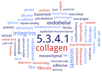
Word Map on EC 5.3.4.1 
-
5.3.4.1
-
collagen
-
laminin
-
integrins
-
endothelial
-
actin
-
fibrosis
-
adhesive
-
basement
-
mesenchymal
-
vessel
-
fiber
-
metastasis
-
proteoglycans
-
vitronectin
-
fibrinogen
-
matrices
-
nephropathy
-
albumin
-
gelatin
-
interstitial
-
mesangial
-
platelet
-
monolayer
-
cytoskeleton
-
glomerular
-
rgd
-
vimentin
-
e-cadherin
-
cell-cell
-
adhesions
-
zeta
-
myofibroblasts
-
willebrand
-
epithelial-mesenchymal
-
factor-beta
-
fak
-
fibrin
-
metalloproteinases
-
preterm
-
dish
-
biomaterials
-
procollagens
-
heparan
-
alpha-smooth
-
dermal
-
tgf-beta
-
plasminogen
-
fibrillar
-
drug development
-
heparin-binding
-
synthesis
-
thrombospondin
-
medicine
-
industry
-
diagnostics
-
pharmacology
-
biotechnology
-
analysis
- 5.3.4.1
- collagen
- laminin
- integrins
- endothelial
- actin
- fibrosis
-
adhesive
-
basement
- mesenchymal
- vessel
- fiber
- metastasis
- proteoglycans
- vitronectin
- fibrinogen
- matrices
- nephropathy
- albumin
- gelatin
-
interstitial
-
mesangial
- platelet
- monolayer
- cytoskeleton
- glomerular
- rgd
- vimentin
- e-cadherin
-
cell-cell
- adhesions
- zeta
- myofibroblasts
- willebrand
-
epithelial-mesenchymal
- factor-beta
- fak
- fibrin
- metalloproteinases
-
preterm
-
dish
-
biomaterials
- procollagens
- heparan
-
alpha-smooth
- dermal
- tgf-beta
- plasminogen
-
fibrillar
- drug development
-
heparin-binding
- synthesis
- thrombospondin
- medicine
- industry
- diagnostics
- pharmacology
- biotechnology
- analysis
Reaction
catalyses the rearrangement of -S-S- bonds in proteins =
Synonyms
5'-MD, 58 kDa glucose regulated protein, 58 kDa microsomal protein, AGR2, anterior gradient homolog 2, BPA-binding protein, CaBP1, CaBP2, Cellular thyroid hormone binding protein, cotyledon-specific chloroplast biogenesis factor CYO1, CYO1, DbsG, disulfide bond isomerase, disulfide bond-forming enzyme, Disulfide interchange enzyme, disulfide isomerase, Disulfide isomerase ER-60, disulfide-bond isomerase, dithiol-disulfide isomerase, Dsb, DsbA, DsbB, DsbC, DsbD, DsbG, ECaSt/PDI, endoplasmic reticulum protein EUG1, Eps1p, ER protein 57, ER58, ER60, ERcalcistorin/protein-disulfide isomerase, ERdj5, Ero1, Erp, ERP-57, ERp-72 homolog, ERp18, ERp27, ERp28, ERp44, Erp46, ERp5, ERp57, ERP59, ERP60, ERp72, Eug1p, fibronectin, gPDI-1, gPDI-2, gPDI-3, HIP-70, HlPDI-1, HlPDI-2, HlPDI-3, Iodothyronine 5'-monodeiodinase, More, Mpd1p, Mpd2p, multifunctional protein disulfide isomerase, ncgl2478, P5, P55, P58, pancreas-specific protein disulfide isomerase, PDI, PDI A4, PDI I, PDI II, pdi-15, PDI-1a, pdi-40, pdi-47, pdi-52, PDI-A, PDI-M, PDI-P5, PDI-related protein, PDI1, PDI11, PDI2, PDI7, PDI8, PDIA1, PDIA2, PDIA3, PDIA4, PDIA6, PDIL-1, PDIL-2, PDIL1-1, PDIL1;1, PDIL1Aalpha, PDIL1B, PDIL2, PDIL2-3, PDIL3A, PDIL4D, PDIL5A, PDILT, PDIp, PDIr, protein disulfide isomerase, protein disulfide isomerase 1, protein disulfide isomerase 2, protein disulfide isomerase 3, protein disulfide isomerase A1, protein disulfide isomerase A3, protein disulfide isomerase A5, protein disulfide isomerase A6, protein disulfide isomerase associated 3, Protein disulfide isomerase P5, protein disulfide isomerase-1, protein disulfide isomerase-11, protein disulfide isomerase-2, protein disulfide isomerase-3, protein disulfide isomerase-8, protein disulfide isomerase-like protein of the testis, protein disulfide isomerase-P5, protein disulfide isomerase-related chaperone Wind, Protein disulfide isomerase-related protein, protein disulfide oxidoreductase, protein disulfide reductase/isomerase, protein disulfide-isomerase A4, Protein disulphide isomerase, Protein ERp-72, protein-disulfide isomerase, R-cognin, RB60, Rearrangease, Reduced ribonuclease reactivating enzyme, Retina cognin, S-S rearrangase, SSO0192, SsPDO, thiol-disulfide oxidoreductase, thiol-protein oxidoreductase, thioredoxin domain-containing protein 5, Thyroid hormone-binding protein, Thyroxine deiodinase, TXNDC5, yPDI


 results (
 results (
 top
top


