6.3.2.3: glutathione synthase
This is an abbreviated version!
For detailed information about glutathione synthase, go to the full flat file.

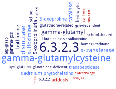
Word Map on EC 6.3.2.3 
-
6.3.2.3
-
gamma-glutamylcysteine
-
dismutase
-
gamma-glutamyl
-
catalase
-
s-transferase
-
sulfoximine
-
cadmium
-
5-oxoprolinuria
-
buthionine
-
5-oxoproline
-
hemolytic
-
acidosis
-
phytochelatins
-
transpeptidase
-
gamma-gcs
-
school
-
school-based
-
pyroglutamic
-
glutathione-related
-
atp-grasp
-
bullied
-
l-buthionine-s,r-sulfoximine
-
juncea
-
6.3.2.2
-
glutathione-deficient
-
homoglutathione
-
gsh-dependent
-
medicine
-
biotechnology
-
synthesis
-
analysis
- 6.3.2.3
- gamma-glutamylcysteine
- dismutase
-
gamma-glutamyl
- catalase
- s-transferase
- sulfoximine
- cadmium
-
5-oxoprolinuria
-
buthionine
- 5-oxoproline
-
hemolytic
- acidosis
- phytochelatins
- transpeptidase
- gamma-gcs
-
school
-
school-based
-
pyroglutamic
-
glutathione-related
-
atp-grasp
-
bullied
-
l-buthionine-s,r-sulfoximine
- juncea
-
6.3.2.2
-
glutathione-deficient
-
homoglutathione
-
gsh-dependent
- medicine
- biotechnology
- synthesis
- analysis
Reaction
Synonyms
Asuc_1947, bifunctional glutathione synthetase, bifunctional GSH synthetase, bifunctional L-glutathione synthetase, gamma -glutamate-cysteine ligase-glutathione synthetase, gamma-GCS, gamma-GCS-GS, gamma-glutamate-cysteine ligase/glutathione synthetase, gamma-glutamylcysteine synthetase-glutathione synthetase, GCL, GCSGS, ghF, glutamate cysteine ligase, glutathione biosynthesis bifunctional protein GshAB, Glutathione synthase, Glutathione synthetase, Glutathione synthetase (tripeptide), GS, GSH synthase, GSH synthetase, GSH-S, GSH2, gshAB, GSHase, gshB, GshF, GshFAp, GshFAs, GSHII, GSHS, GSHS1, GSS, hGS, L-glutathione synthetase, More, Phytochelatin synthetase, StGCL-GS, Synthetase, glutathione, TAGS1, TaGS2, TbGS, ZmGS


 results (
 results (
 top
top


