6.1.1.22: asparagine-tRNA ligase
This is an abbreviated version!
For detailed information about asparagine-tRNA ligase, go to the full flat file.

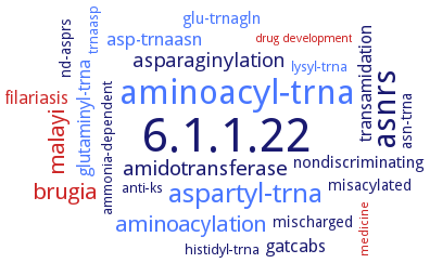
Word Map on EC 6.1.1.22 
-
6.1.1.22
-
aminoacyl-trna
-
asnrs
-
aspartyl-trna
-
malayi
-
brugia
-
aminoacylation
-
amidotransferase
-
asparaginylation
-
asp-trnaasn
-
glutaminyl-trna
-
gatcabs
-
filariasis
-
transamidation
-
nondiscriminating
-
glu-trnagln
-
mischarged
-
misacylated
-
asn-trna
-
nd-asprs
-
ammonia-dependent
-
anti-ks
-
lysyl-trna
-
trnaasp
-
histidyl-trna
-
medicine
-
drug development
- 6.1.1.22
- aminoacyl-trna
- asnrs
- aspartyl-trna
- malayi
- brugia
- aminoacylation
-
amidotransferase
-
asparaginylation
- asp-trnaasn
- glutaminyl-trna
- gatcabs
- filariasis
-
transamidation
-
nondiscriminating
- glu-trnagln
-
mischarged
-
misacylated
-
asn-trna
- nd-asprs
-
ammonia-dependent
-
anti-ks
- lysyl-trna
- trnaasp
-
histidyl-trna
- medicine
- drug development
Reaction
Synonyms
AS-AR, AsnRS, Asparagine synthetase A, Asparagine translase, asparagine tRNA synthetase, Asparagine--tRNA ligase, Asparaginyl transfer ribonucleic acid synthetase, Asparaginyl transfer RNA synthetase, asparaginyl tRNA synthetase, Asparaginyl-transfer ribonucleate synthetase, asparaginyl-transfer RNA synthetase, Asparaginyl-tRNA synthetase, Asparagyl-transfer RNA synthetase, class IIb asparaginyl-tRNA synthetase, More, NARS, NARS2, NRS, Potentially protective 63 kDa antigen, PYRAB02460, Synthetase, asparaginyl-transfer ribonucleate
ECTree
Advanced search results
Crystallization
Crystallization on EC 6.1.1.22 - asparagine-tRNA ligase
Please wait a moment until all data is loaded. This message will disappear when all data is loaded.
identification of peptidyl regions that are surface-accessible and available for antibody binding
purified apo-AsnRS, AsnRS in complex with AsnAMS, and in complex with Mg2+, ATP, and l-Asp-beta-NOH, X-ray diffraction structure determination and analysis at 1.9-2.4 A resolution
-
purified dimeric enzyme with bound Mg2+ and a non-hydrolyzable analogue of asparaginyl adenylate, ASNAMS, X-ray diffraction structure determination and analysis at 1.9 A resolution, modeling
-
sitting drop vapor diffusion method, using 100 mM Tris-HCl (pH 7.0), 0.2 M NaCl, and 32% (w/v) polyethylene glycol 3350
AsnRS complexed with asparaginyl-adenylate Asn-AMP, or free AsnRS and AsnRS complexed with an Asn-AMP analog Asn-SA, sitting-drop vapor diffusion method at 20°C, 0.001 ml protein solution is mixed with an equal volume of reservoir solution containing 25 mM sodium cacodylate buffer, pH 6.5, 50 mM sodium acetate trihydrate, and 7.5% w/v PEG 8000, equilibration against 0.5 ml reservoir solution, for ligand complex formation the crystals are soaked in a reservoir solution, containing 1.5 mM AMP-PNP, 1.5 mM asparagine, and 2 mM MgCl2, for approximately 10 h, followed by a reservoir solution containing 25% 2-methyl-2,4-pentanediol for a few seconds, X-ray diffraction strucure determination and analysis at 1.45 A, and 1.98 A and 1.8 A resolution, respectively
AsnRS with bound asparaginyladenylate, AsnAMP, X-ray diffraction structure anaylsis at 2.6 A resolution
-
recombinant enzyme, native or complexed withATP and asparaginyl-adenylate, 3 different crystal forms, X-ray diffraction structure determination at 2.6 A resolution and analysis


 results (
 results (
 top
top


