6.1.1.14: glycine-tRNA ligase
This is an abbreviated version!
For detailed information about glycine-tRNA ligase, go to the full flat file.

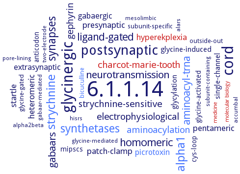
Word Map on EC 6.1.1.14 
-
6.1.1.14
-
cord
-
glycinergic
-
postsynaptic
-
strychnine
-
alpha1
-
synthetases
-
synapses
-
ligand-gated
-
aminoacyl-trna
-
homomeric
-
gabaars
-
neurotransmission
-
electrophysiological
-
aminoacylation
-
charcot-marie-tooth
-
strychnine-sensitive
-
gephyrin
-
patch-clamp
-
hyperekplexia
-
presynaptic
-
heteromeric
-
startle
-
pentameric
-
gabaergic
-
extrasynaptic
-
picrotoxin
-
glycylation
-
glycine-induced
-
single-channel
-
glycine-activated
-
cys-loop
-
anticodon
-
mipscs
-
subunit-specific
-
bicuculline
-
glycine-gated
-
outside-out
-
subunit-containing
-
alars
-
two-electrode
-
alpha2beta
-
mesolimbic
-
pore-lining
-
hisrs
-
glycine-mediated
-
accumbal
-
gabaar-mediated
-
molecular biology
-
medicine
- 6.1.1.14
- cord
-
glycinergic
-
postsynaptic
- strychnine
- alpha1
- synthetases
-
synapses
-
ligand-gated
- aminoacyl-trna
-
homomeric
-
gabaars
-
neurotransmission
-
electrophysiological
- aminoacylation
- charcot-marie-tooth
-
strychnine-sensitive
-
gephyrin
-
patch-clamp
- hyperekplexia
-
presynaptic
-
heteromeric
-
startle
-
pentameric
-
gabaergic
-
extrasynaptic
- picrotoxin
-
glycylation
-
glycine-induced
-
single-channel
-
glycine-activated
-
cys-loop
-
anticodon
-
mipscs
-
subunit-specific
- bicuculline
-
glycine-gated
-
outside-out
-
subunit-containing
- alars
-
two-electrode
-
alpha2beta
-
mesolimbic
-
pore-lining
- hisrs
-
glycine-mediated
-
accumbal
-
gabaar-mediated
- molecular biology
- medicine
Reaction
Synonyms
GARS, Glycine--tRNA ligase, Glycyl translase, glycyl tRNA synthetase, Glycyl-transfer ribonucleate synthetase, Glycyl-transfer ribonucleic acid synthetase, Glycyl-transfer RNA synthetase, Glycyl-tRNA synthetase, glycyl-tRNA synthetase 1, GlyRS, GlyRS1, GlyRS2, GRS1, More, Synthetase, glycyl-transfer ribonucleate
ECTree
Advanced search results
Crystallization
Crystallization on EC 6.1.1.14 - glycine-tRNA ligase
Please wait a moment until all data is loaded. This message will disappear when all data is loaded.
alpha-subunit the enzyme and its complexes with ATP, and ATP and glycine, sitting drop vapor diffusion method, using 0.1 M Bis-Tris propane:NaOH pH 7.0 and 0.7 M magnesium formate (complex with glycine), or 0.1 M Tris-HCl pH 8.5 and 0.3 M magnesium chloride (complex with ATP), or 0.1 M Na2HPO4-citrate acid pH 4.2, 0.2 M lithium sulfate and 20% (w/v) PEG 1000 (complex with ATP and glycine)
apo form of full-length E71G mutant and tRNA-bound mutant complex E71G/C157R, vapor diffusion method, using 13-15% (w/v) PEG 6000, 0.1 M sodium citrate buffer (pH 5.5), and 0.1 M NaCl
purified His-tagged wild-type and S581L mutant enzymes, sitting drop vapour diffusion nanocrystallization method, mutant S581L GlyRS from 20% PEG 3350, 0.2 M Na2SO4, 0.1 M Bis-Tris propane, pH 6.5, wild-type GlyRS from 20% PEG 3350, 0.2M sodium bromide, 0.1 M bis-Tris propane pH 8.5, X-ray diffraction structure determination and anaylsis at 2.8A and 3.1 A resolution, respectively
-
purified recombinant soluble enzyme, hanging drop vapour diffusion method, 0.001 ml of 8 mg/ml protein in 10 mM HEPES, pH 7.0, with 20 mM NaCl is mixed with 0.001 ml of reservoir solution containing 10% PEG 6000, 0.1 HEPES pH 6.5, 0.01 Tris-HCl, pH 8.5, 0.5 NaCl and 0.1 magnesium acetate at room temperature, 2 days, X-ray diffraction structure determination and analysis at 3.0 A resolution
vapor diffusion method, using 0.2% (w/v) 4-diaminobutane, 0.2% (w/v) cystamine dihydrochloride, 0.2% (w/v) diloxanide furoate, 0.2% (w/v) sarcosine, 0.2% (w/v) spermine, and 0.02 M HEPES sodium (pH 6.8) at a 2:1:1 ratio


 results (
 results (
 top
top


