4.98.1.1: protoporphyrin ferrochelatase
This is an abbreviated version!
For detailed information about protoporphyrin ferrochelatase, go to the full flat file.

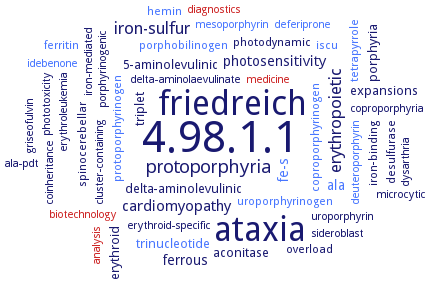
Word Map on EC 4.98.1.1 
-
4.98.1.1
-
ataxia
-
friedreich
-
protoporphyria
-
erythropoietic
-
iron-sulfur
-
cardiomyopathy
-
fe-s
-
ferrous
-
ala
-
photosensitivity
-
aconitase
-
porphyria
-
expansions
-
delta-aminolevulinic
-
trinucleotide
-
triplet
-
5-aminolevulinic
-
erythroid
-
coproporphyrinogen
-
iron-binding
-
spinocerebellar
-
overload
-
photodynamic
-
hemin
-
iscu
-
desulfurase
-
ferritin
-
protoporphyrinogen
-
porphobilinogen
-
tetrapyrrole
-
uroporphyrinogen
-
diagnostics
-
biotechnology
-
griseofulvin
-
sideroblast
-
coproporphyria
-
iron-mediated
-
uroporphyrin
-
analysis
-
dysarthria
-
cluster-containing
-
phototoxicity
-
microcytic
-
porphyrinogenic
-
deferiprone
-
erythroid-specific
-
coinheritance
-
erythroleukemia
-
ala-pdt
-
medicine
-
idebenone
-
deuteroporphyrin
-
mesoporphyrin
-
delta-aminolaevulinate
- 4.98.1.1
-
ataxia
-
friedreich
-
protoporphyria
-
erythropoietic
-
iron-sulfur
-
cardiomyopathy
- fe-s
-
ferrous
- ala
-
photosensitivity
- aconitase
-
porphyria
-
expansions
-
delta-aminolevulinic
- trinucleotide
-
triplet
-
5-aminolevulinic
-
erythroid
- coproporphyrinogen
-
iron-binding
-
spinocerebellar
-
overload
-
photodynamic
- hemin
- iscu
-
desulfurase
- ferritin
- protoporphyrinogen
- porphobilinogen
- tetrapyrrole
- uroporphyrinogen
- diagnostics
- biotechnology
-
griseofulvin
-
sideroblast
-
coproporphyria
-
iron-mediated
-
uroporphyrin
- analysis
-
dysarthria
-
cluster-containing
-
phototoxicity
-
microcytic
-
porphyrinogenic
- deferiprone
-
erythroid-specific
-
coinheritance
-
erythroleukemia
-
ala-pdt
- medicine
- idebenone
- deuteroporphyrin
- mesoporphyrin
-
delta-aminolaevulinate
Reaction
Synonyms
chelatase, ferro-, EC 4.99.1.1, FC1, FC2, FeC, FeCH, ferro-protoporphyrin chelatase, ferrochelatase, ferrochelatase 1, ferrochelatase I, ferrochelatase II, frataxin, HEM15, heme synthase, heme synthetase, hemH, HemH1, HemH2, hFC, host red cell ferrochelatase, iron chelatase, parasite genome-coded ferrochelatase, PfFC, PPIX ferrochelatase, protohaem ferrolyase, protoheme ferro-lyase, protoheme ferrolyase, protoheme lyase, protoporhyrin IX ferrochelatase, protoporphyrin (IX) ferrochelatase, protoporphyrin IX ferrochelatase, Shew_1140, Shew_2229, type II ferrochelatase
ECTree
Advanced search results
Crystallization
Crystallization on EC 4.98.1.1 - protoporphyrin ferrochelatase
Please wait a moment until all data is loaded. This message will disappear when all data is loaded.
crystals of wild-type and mutants H183A, H88A and K87A with iron, by addition of solid (NH4)2Fe(SO4)2 x 6 H2O, 1.2 A to 1.7 A resolution
Mg2+ is required for the formation of Bacillus subtilis ferrochelatase crystals
overview
structure in presence of iron. Only a single iron ion is found in the active site, coordinated in a square pyramidal fashion by two amino acid residues, His183 and Glu264, and three water molecules. This iron ion is not present in the structure of a His183Ala modified ferrochelatase. Insertion of a metal ion into protoporphyrin IX by ferrochelatase occurs from a metal binding site represented by His183 and Glu264
by the hanging drop method, crystals of mutant E343K with the substrate protoporphyrin IX and mutant R115L without bound substrate. Enzyme with porphyrin bound possesses a significantly more closed active site conformation. In the substrate-bound form, the jaws of the active site mouth are closed so that the porphyrin substrate is completely engulfed in the pocket
crystals of wild-type enzyme in the presence of ammonium chloride or manganese chloride, wild-type enzyme adopts a predominantly open conformation
-
mutant enzymes E343D and E343Q in the presence of cobalt chloride, hanging drop vapor diffusion method, using 0.1 M Bis-Tris pH 6.5, 0.05 M ammonium sulfate and a range of pentaerythritol ethoxylate (15/4 EO/OH) varying from 20% to 35% (w/v)
quantum mechanical/molecular mechanics studies. The ferrous iron probably coordinates with residue Met76 at the binding site, and His263 plays the role of proton acceptor. The rate-determining step is either the first proton removed by His263 or the proton transition within the porphyrin with an energy barrier of 14.99 or 14.87 kcal/mol, respectively
vapor diffusion hanging drop, 291 K, 0.05 M ammonium sulfate, 0.1 M Bis-Tris pH 6.5, 20% pentaerythritolethyloxylate, data for crystals obtained with wild-type human ferrochelatase treated with protoporphyrin IX and Hg, 1.6 A resolution; data for crystals obtained with wild-type human ferrochelatase treated with protoporphyrin IX and Cd, 1.8 A resolution, data for crystals obtained with the F110A variant of human ferrochelatase treated with deuteroporphyrin and Mn, 2.0 A resolution, data for crystals obtained with the F110A variant of human ferrochelatase treated with deuteroporphyrin and Ni, 2.20 A resolution
free form, in complex with Co(II) and in complex with Cd(II) and Hg(I)


 results (
 results (
 top
top


