3.5.2.3: dihydroorotase
This is an abbreviated version!
For detailed information about dihydroorotase, go to the full flat file.

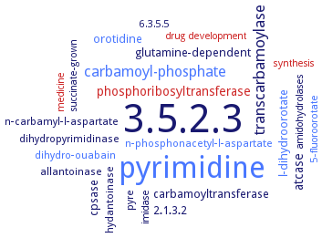
Word Map on EC 3.5.2.3 
-
3.5.2.3
-
pyrimidine
-
transcarbamoylase
-
carbamoyl-phosphate
-
phosphoribosyltransferase
-
atcase
-
orotidine
-
carbamoyltransferase
-
glutamine-dependent
-
l-dihydroorotate
-
2.1.3.2
-
cpsase
-
pyre
-
dihydropyrimidinase
-
hydantoinase
-
dihydro-ouabain
-
n-phosphonacetyl-l-aspartate
-
n-carbamyl-l-aspartate
-
allantoinase
-
imidase
-
synthesis
-
succinate-grown
-
amidohydrolases
-
6.3.5.5
-
drug development
-
medicine
-
5-fluoroorotate
- 3.5.2.3
- pyrimidine
-
transcarbamoylase
- carbamoyl-phosphate
- phosphoribosyltransferase
- atcase
- orotidine
-
carbamoyltransferase
-
glutamine-dependent
- l-dihydroorotate
-
2.1.3.2
- cpsase
-
pyre
- dihydropyrimidinase
- hydantoinase
- dihydro-ouabain
- n-phosphonacetyl-l-aspartate
-
n-carbamyl-l-aspartate
- allantoinase
- imidase
- synthesis
-
succinate-grown
-
amidohydrolases
-
6.3.5.5
- drug development
- medicine
- 5-fluoroorotate
Reaction
Synonyms
amidohydrolase family protein, BcDHOase, CAD, carbamoylaspartic dehydrase, Class I DHOase, DHO, DHOase, dihydroorotase, dihydroorotase domain, dihydroorotate dehydrolase, hDHOase, huDHOase, human DHOase domain, LdDHOase, More, pyrC, type I DHOase, type II DHO, VcDHO, YpDHO
ECTree
Advanced search results
Crystallization
Crystallization on EC 3.5.2.3 - dihydroorotase
Please wait a moment until all data is loaded. This message will disappear when all data is loaded.
crystal structure analysis of the zinc environment at ambient pressure, at 600 bar, and at 1200 bar
hanging drop method, monoclinic structure has a novel cysteine ligand to the zinc that blocks the active site and functions as a cysteine switch, active site residues are located in disordered loops, which may function as a disorder-to-order entropy switch
purified recombinant detagged wild-type enzyme complexed with N-carbamoyl-L-aspartate or (S)-dihydroorotate, hanging drop vapor diffusion method, mixing of 10 mg/ml protein solution, containing 1.5 mM ligand, with reservoir solution containing 0.1 M Bis-Tris, pH 5.5, 0.2 M NaCl, and 20% PEG 3350, X-ray diffraction structure determination and analysis at 2.45 A resolution, chains A and B are in one asymmetric unit. The active site of each monomer, and both substrates have relatively weak electron density due to low substrate occupancy
to 2.6 A resolution. The overall tertiary structure formed from the TIM-barrel secondary structure motif is conical, with the active site residing at the base of the cone. The active site includes a binuclear Zn center, with two histidine (His59 and His61) and two asparagine (Asp151 and Asp304) residues bound to the more buried first Zn atom and two histidine (178 and231) residues and Asp151 bound to the second Zn atom. The carboxylate side group of Asp151 forms a bridge between the two Zn atoms
crystallized without ligand and in the presence of the inhibitors 2-oxo-1,2,3,6-tetrahydropyrimidine-4,6-dicarboxylate and 5-fluoroorotate, hanging drop vapour diffusion method with 15-20% (w/v) polyethylene glycol 3350, 0.1 M MES (pH 6.0-6.5), 75 mM MgCl2, 150 mM KCl (with ligand) or 20-25% polyethylene glycol 3350, 0.1 M Na HEPES (pH 7) and 0.2 M NaF (without ligand)
hanging drop vapour diffusion method with 15-20% polyethylene glycol 3350, 0.1 M MES, pH 6.0-6.5, 75 mM MgCl2, and 150 mM KCl
in complex with 5-fluoroorotate, hanging drop vapour diffusion method 14-16% PEG 3350, 0.1 M MES pH 6.25, 25 mM MgCl2, 0.2 M KCl and 30% sucrose
in the presence of enantiomerically pure L-dihydroorotate, refined at 1.9 Angstrom resolution
human DHOase domain K1556A mutant, hanging drop vapor diffusion method, mixing of 20 mg/ml protein in 20 mM HEPES and 100 mM NaCl, pH 7.0, with reservoir solution containing 2 M sodium chloride, 100 mM MES, 200 mM sodium acetate, pH 6.5, at room temperature, X-ray diffraction structure determination and analysis at 2.77 A resolution
several F1563 mutant variants of huDHOase bound to dihydroorotate, X-ray diffraction structure determination and analysis at 1.46-2.12 A resolution
purified recombinant His-tagged enzyme, enzyme proteolysis before crystallization, sitting drop vapor diffusion technique, mixing of 0.001 ml of 14 mg/ml protein in 20 mM HEPES, pH 7.5, 150 mM NaCl, 10% glycerol, 0.1% sodium azide, 0.5 mM TCEP, and and 20 mM L-citrulline, with 0.0015 ml of crystallization solution containing 133 mM sodium acetate, 67 mM sodium cacodylate-HCl, pH 6.5, 20% w/v PEG 8000, and 2.3% v/v 1-butanol, and equilibration against 0.034 ml of reservoir solution of 1.5 M NaCl, 4-7 days, 16°C, X-ray diffraction structure determination and analysis at 1.95 A resolution, molecular replacement, modelling
purified recombinant His-tagged enzyme, sitting drop vapor diffusion technique, mixing of 400 nl of 10 mg/ml protein in 20 mM Tris-HCl, pH 7.5, 150 mM NaCl, 10% glycerol, 0.1% sodium azide, and 0.5 mM TCEP, with 400 nl of crystallization solution containing 0.15 M DL-malic acid, pH 7.0, and 20% w/v PEG 3350, equilibration against 0.034 ml of reservoir solution of 1.3 M NaCl, 4-7 days, 16°C, X-ray diffraction structure determination and analysis at 2.4 A resolution, molecular replacement, modelling


 results (
 results (
 top
top


