3.5.1.2: glutaminase
This is an abbreviated version!
For detailed information about glutaminase, go to the full flat file.
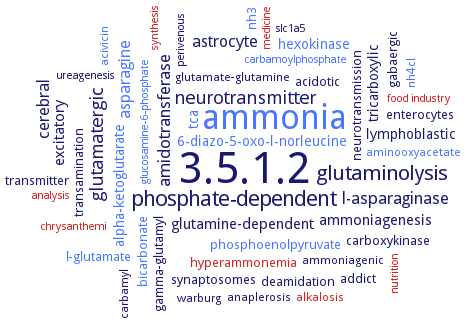
Word Map on EC 3.5.1.2
-
3.5.1.2
-
ammonia
-
phosphate-dependent
-
glutaminolysis
-
neurotransmitter
-
glutamatergic
-
l-asparaginase
-
amidotransferase
-
cerebral
-
astrocyte
-
asparagine
-
excitatory
-
ammoniagenesis
-
glutamine-dependent
-
6-diazo-5-oxo-l-norleucine
-
hexokinase
-
tricarboxylic
-
tca
-
alpha-ketoglutarate
-
lymphoblastic
-
phosphoenolpyruvate
-
deamidation
-
gamma-glutamyl
-
carboxykinase
-
synaptosomes
-
bicarbonate
-
neurotransmission
-
enterocytes
-
addict
-
gabaergic
-
transamination
-
transmitter
-
l-glutamate
-
acidotic
-
hyperammonemia
-
nh3
-
carbamyl
-
warburg
-
ammoniagenic
-
anaplerosis
-
aminooxyacetate
-
alkalosis
-
nh4cl
-
acivicin
-
glutamate-glutamine
-
perivenous
-
slc1a5
-
chrysanthemi
-
carbamoylphosphate
-
ureagenesis
-
analysis
-
medicine
-
nutrition
-
synthesis
-
food industry
-
glucosamine-6-phosphate
- 3.5.1.2
- ammonia
-
phosphate-dependent
-
glutaminolysis
-
neurotransmitter
-
glutamatergic
- l-asparaginase
-
amidotransferase
- cerebral
- astrocyte
- asparagine
-
excitatory
-
ammoniagenesis
-
glutamine-dependent
- 6-diazo-5-oxo-l-norleucine
- hexokinase
-
tricarboxylic
- tca
- alpha-ketoglutarate
- lymphoblastic
- phosphoenolpyruvate
-
deamidation
-
gamma-glutamyl
-
carboxykinase
-
synaptosomes
- bicarbonate
-
neurotransmission
- enterocytes
-
addict
-
gabaergic
-
transamination
-
transmitter
- l-glutamate
-
acidotic
- hyperammonemia
- nh3
-
carbamyl
-
warburg
-
ammoniagenic
-
anaplerosis
- aminooxyacetate
- alkalosis
- nh4cl
- acivicin
-
glutamate-glutamine
-
perivenous
-
slc1a5
- chrysanthemi
- carbamoylphosphate
-
ureagenesis
- analysis
- medicine
- nutrition
- synthesis
- food industry
- glucosamine-6-phosphate
Reaction
Synonyms
AnsB, AoGls, GA, GAB, GAC, GahB, GLA, GLNase, GLS, GLS1, GLS2, GlsA, glutaminase, glutaminase 1, glutaminase 2, glutaminase A, glutaminase B, glutaminase C, glutaminase I, glutaminase K, glutaminase L, glutaminase-1, glutaminase-2, glutaminase-B, glutamine amidohydrolase, glutamine aminohydrolase, glutamine deamidating enzyme, K-glutaminase, KAG, KGA, kidney-type glutaminase, kidney-type-glutaminase, L-glutaminase, L-glutamine amidohydrolase, LAG, LGA, liver-type glitaminase, liver-type glutaminase, Mglu, Micrococcus glutaminase Mglu, Micrococcus luteus K-3-type glutaminase, mitochondrial glutaminase, N-PAG, neuroblastoma glutaminase, neuroblastoma PAG, Nit 2, nitrilase 2, omega-amidase, PAG, PDX2, phosphate activated glutaminase, phosphate-activated glutaminase, phosphate-activated L-glutamine amidohydrolase, salt-tolerant glutaminase, YaaE
ECTree

Advanced search results
Crystallization
Crystallization on EC 3.5.1.2 - glutaminase
 top
top
