3.2.1.86: 6-phospho-beta-glucosidase
This is an abbreviated version!
For detailed information about 6-phospho-beta-glucosidase, go to the full flat file.

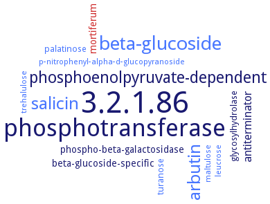
Word Map on EC 3.2.1.86 
-
3.2.1.86
-
phosphotransferase
-
beta-glucoside
-
arbutin
-
salicin
-
phosphoenolpyruvate-dependent
-
antiterminator
-
beta-glucoside-specific
-
mortiferum
-
phospho-beta-galactosidase
-
turanose
-
palatinose
-
glycosylhydrolase
-
trehalulose
-
leucrose
-
p-nitrophenyl-alpha-d-glucopyranoside
-
maltulose
- 3.2.1.86
-
phosphotransferase
- beta-glucoside
- arbutin
- salicin
-
phosphoenolpyruvate-dependent
-
antiterminator
-
beta-glucoside-specific
- mortiferum
- phospho-beta-galactosidase
- turanose
- palatinose
-
glycosylhydrolase
- trehalulose
- leucrose
- p-nitrophenyl-alpha-d-glucopyranoside
- maltulose
Reaction
Synonyms
6-phospho-alpha-glucosidase, 6-phospho-beta-glucosidase, AscB protein, bgl-2, BglA, BglA-2, BglA3, bglD, BglT, CelD, Cellobiose-6-phosphate hydrolase, Gan1D, LacG1, LacG2, More, P-beta-glc, PalH, Pbgl25-217, phospho-alpha-glucosidase, phospho-beta-glucosidase, phospho-beta-glucosidase A, phosphocellobiase, SPD_0247, SPy1599
ECTree
Advanced search results
Crystallization
Crystallization on EC 3.2.1.86 - 6-phospho-beta-glucosidase
Please wait a moment until all data is loaded. This message will disappear when all data is loaded.
in complex with cellobiose-6-phosphate, 6-phospho-D-galactose or 6-phospho-D-glucose, hanging drop vapor diffusion method, using 16-22% (w/v) polyethylene glycol 8 K, 3% (w/v) methylpentanediol, 0.1 M imidazole buffer pH 6.5
purified recombinant enzyme in apoform or complexed with thiocellobiose 6-phosphate, sitting drop vapour diffusion method, mixing of 0.001 ml of 10 mg/ml protein in 20 mM Tris-Cl, pH 8.0, 100 mM NaCl with 0.001 ml of reservoir solution containing 15% PEG 5000MME, and 0.1 M sodium citrate, pH 5.6, 16°C, X-ray diffraction structure determination and analysis at 2.0-2.4 A resolution, molecular replacement method
crystals with C2, P2 twinned, and P2 not twinned structures, hanging-drop vapour diffusion method, mixing equal volumes of 20 mg/ml protein with 0.1 M Bis-Tris propane, pH 7.5, 0.2 M sodium fluoride or sodium bromide, 20% PEG 3350 at 19°C, X-ray diffraction structure determination and analysis at 2.51-2.61 A resolution, three-dimensional structure analysis of the enzyme in a number of crystal forms complicated by unusual crystallographic twinning, the enzyme shows a classical (beta/alpha)8-barrel structure, molecular replacement method
in complex with 6-phospho-cyclophellitol, sitting drop vapor diffusion method, using 20% (w/v) PEG3350, NaBr 0.15 M, Bis-Tris propane 0.1 M, pH 7.5
hanging-drop vapor-diffusion method, crystal structure of the native selenomethionine-substituted enzyme (2.85 A resolution) and of the enzyme in complex with NAD+, Mn2+ and glucose 6-phosphate (2.55 A resolution)


 results (
 results (
 top
top


