3.2.1.18: exo-alpha-sialidase
This is an abbreviated version!
For detailed information about exo-alpha-sialidase, go to the full flat file.
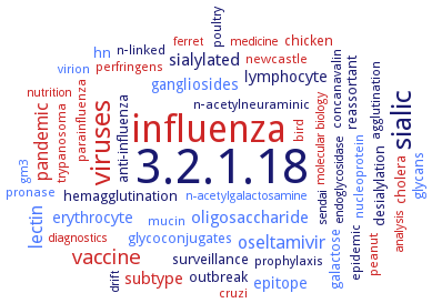
Word Map on EC 3.2.1.18
-
3.2.1.18
-
influenza
-
sialic
-
viruses
-
vaccine
-
pandemic
-
oseltamivir
-
lectin
-
sialylated
-
oligosaccharide
-
subtype
-
epitope
-
erythrocyte
-
gangliosides
-
lymphocyte
-
reassortant
-
hemagglutination
-
anti-influenza
-
chicken
-
glycans
-
surveillance
-
galactose
-
cholera
-
desialylation
-
hn
-
glycoconjugates
-
outbreak
-
cruzi
-
poultry
-
virion
-
bird
-
pronase
-
parainfluenza
-
epidemic
-
n-acetylneuraminic
-
trypanosoma
-
peanut
-
mucin
-
n-linked
-
newcastle
-
agglutination
-
perfringens
-
nucleoprotein
-
prophylaxis
-
concanavalin
-
medicine
-
drift
-
sendai
-
analysis
-
n-acetylgalactosamine
-
ferret
-
nutrition
-
endoglycosidase
-
molecular biology
-
diagnostics
-
gm3
- 3.2.1.18
- influenza
-
sialic
- viruses
- vaccine
- pandemic
- oseltamivir
- lectin
-
sialylated
- oligosaccharide
- subtype
- epitope
- erythrocyte
- gangliosides
- lymphocyte
-
reassortant
-
hemagglutination
-
anti-influenza
- chicken
- glycans
-
surveillance
- galactose
- cholera
-
desialylation
- hn
- glycoconjugates
-
outbreak
- cruzi
-
poultry
- virion
- bird
- pronase
- parainfluenza
-
epidemic
-
n-acetylneuraminic
- trypanosoma
- peanut
- mucin
-
n-linked
- newcastle
-
agglutination
- perfringens
- nucleoprotein
-
prophylaxis
-
concanavalin
- medicine
-
drift
-
sendai
- analysis
- n-acetylgalactosamine
- ferret
- nutrition
-
endoglycosidase
- molecular biology
- diagnostics
- gm3
Reaction
Synonyms
acetylneuraminidase, Acetylneuraminyl hydrolase, acid sialidase, acylneuraminyl glycohydrolase, alpha-N-acylneuraminate glycohydrolase, alpha-neuraminidase, alpha-sialidase, alpha2,6-sialidase, alpha2,6-sialyltransferase, alpha2,6-trans-sialidase, Am0705, Am0707, Am1757, Am2085, Cytosolic sialidase, DELTA15Pd2,6ST, endo-N, endo-N-acylneuraminidase, endo-sialidase, endoNF, endosialidase, exo-alpha-sialidase, exo-sialidase, G9 sialidase, Ganglioside sialidase, ganglioside-specific sialidase, GH33C, glycosyl hydrolase, haemagglutinin-neuraminidase protein, hemagglutinin-neuraminidase, hemagglutinin-neuraminidase glycoprotein, HN, HsNEU2, IID sialidase, KDNase, Lysosomal sialidase, Major 85 kDa surface antigen, Major surface antigen, Membrane sialidase, MmNEU3, More, Mouse skeletal muscle sialidase, MSS, MTS, mucopolysaccharide N-acetylneuraminylhydrolase, Murine thymic sialidase, N-acetyl-alpha-neuraminidase, N-acetyl-alpha-neuraminidase 1, N-acetyl-alpha-neuraminidase 2, N-acetyl-alpha-neuraminidase 3, N-acetyl-alpha-neuraminidase 4, N-acetylneuraminosyl glycohydrolase, N-acylneuraminate glycohydrolase, N-acylneuraminosyl glycohydrolase, NA, NA1, NA2, NAN1, NanA, NANase, NanB, NanC, NanH, NanI, NanICD, NanJ, NanPs, Neu, NEU1, NEU2, Neu3, Neu3a, Neu3d, Neu3e, NEU4, NEU4 long, NEU4 short, NEU4L, Neu4L sialidase, neuramindase, neuraminidase, neuraminidase 1, neuraminidase 2, neuraminidase 3, neuraminidase-1, PA2794, SA85-1.1 protein, SA85-1.2 protein, SA85-1.3 protein, SiaBb1, sialidase, sialidase 4, sialidase II, sialidase Neu2, sialidase-2, sialidase-3, sialidase-4, sialidase/neuraminidase mutant F129A, sialyl hydrolase, STNA, TcTS, TDE0471, Tr6, trans-sialidase, trans-sialidase 1, TSia, VCNA
ECTree

Advanced search results
Crystallization
Crystallization on EC 3.2.1.18 - exo-alpha-sialidase
Please wait a moment until all data is loaded. This message will disappear when all data is loaded.
 top
topCRYSTALLIZATION (Commentary) 

ORGANISM
UNIPROT
LITERATURE
sitting drop vapor diffusion method, using 20% (w/v) polyethylene glycol 4000, 0.2 M ammonium citrate tri-basic (pH 7.0) and 0.1 M Tris pH 8.5
catalytic domain of isoform NanI, to 0.97 A resolution, and complexes with its substrate sialic acid to 0.97A resolution, with transition-state analogue 2-deoxy-2,3-dehydro-N-acetylneuraminic acid to 1.5 A resolution, and with a covalent intermediate formed using a fluorinated sialic acid analogue to 1.2 A resolution
crystallization of the catalytic domain by sitting-drop vapour-diffusion. Crystals belong to space group P2(1)2(1)2(1) with unit-cell parameters a = 96.98, b = 69.41, c = 72.69 A and one monomer per asymmetric unit. The crystals diffract to at least 0.92 A
-
in complex with 2-(cyclohexylamino)ethanesulfonic acid, hanging drop vapor diffusion method, using 20% (w/v) PEG 3350 and 0.2 M ammonium sulfate
-
sitting drop vapor diffusion method, using 0.1 M MES monohydrate pH 6.0 and 14% (w/v) polyethylene glycol 4000
sitting drop vapor diffusion method, using 5% (v/v) 2-methyl-2,4-pentanediol, 0.1 M HEPES pH 7.5 and 10% (w/v) polyethylene glycol 10000
hanging drop method, high resolution X-ray structures of human sialidase Neu2, in its apo form and in complex with the inhibitor 2-deoxy-2,3-dehydro-N-acetylneuraminic acid. The structure shows the canonical six-blade beta-propeller observed in viral and bacterial sialidases with its active site in a shallow crevice. In the complex structure, the inhibitor lies in the catalytic crevice surrounded by ten amino acids
modeling of three-dimensional structures of NEU1, NEU3 and NEU4 based on the crystal structure of NEU2 using a homology modeling program
modeling of three-dimensional structures of NEU3 based on the crystal structure of NEU2 using a homology modeling program
modeling of three-dimensional structures of NEU4 based on the crystal structure of NEU2 using a homology modeling program
mutant enzymes in complex with oseltamivir carboxylate, hanging drop vapor diffusion method, mutant I223V: 0.1 M HEPES pH 6.75, 7% (w/v) PEG 8000, mutant S247N: 0.1 M HEPES pH 6.7, 8.5% (w/v) PEG 8000, mutant H275Y: 0.1 M HEPES pH 7.0, 8% (w/v) PEG 8000, mutant I223V/H275Y: 0.1 M HEPES pH 7.0, 7% (w/v) PEG 8000, and mutant S247N/H275Y: 0.1 M HEPES pH 7.5, 5% (w/v) PEG 8000
crystallization of the enzyme alone or in complex with sialic acid, modeling
free enzyme, sitting drop vapor diffusion method, using 100 mM HEPES (pH 7.0), 5% tacsimate, 7% (w/v) PEG 5000MME, at 21°C
-
hanging drop vapor diffusion method, using 20% PEG 6K and 0.1 M bicine, pH 5.0
-
56500 Da domain that retains full enzymatic activity, in presence of inhibitor 2-deoxy-2,3-dehydro-N-acetyl neuraminic acid. 2.5 A resolution, space group P212121
free enzyme and in complex with N-acetylneuraminic acid or 2,3-dehydro-2-deoxy-N-acetylneuraminic acid, sitting drop vapor diffusion method, using 100 mM HEPES (pH 7.0) and 30% Jeffamine ED-2001 (pH 7.0), at 21°C
-
comparison of the catalytic cleft plasticity of free and ligand-bound forms of Trypanosoma rangeli sialidase and Trypanosoma cruzi trans-sialidase using molecular dynamics simulations. The Trypanosoma cruzi enzyme has a very flexible, widely open catalytic cleft, mostly due to resiude W312 loop motion, in apo form. In ligan-bound form, the flexibility and solvent exposure is significantly reduced. The Trypanosoma rangeli sialidase maintains a more open catalytic cleft in both apo and holo forms
-
wild-type and mutant D59A, complexed with 2 mM 3-deoxy-N-acetylneuraminic acid to give a covalent intermediate, in 2 M ammonium sulfate, 100 mM HEPES, pH 8.0, 2%PEG 400, used for microseeding, in 10% PEG 4000, 100 mM Tris-HCl, pH 7.5, and 5% isopropanol, soaking in buffer with 5 mM 2,3-difluoro-sialic acid at 25°C, soaking in buffer containing 10 mM alpha-(2-3)-sialyllactose or 2'(4-methylumbelliferyl)-alpha-D-N-acetylneuraminic acid, freezing and X-ray diffraction structure determination and analysis at 1.6 A
-
comparison of the catalytic cleft plasticity of free and ligand-bound forms of Trypanosoma rangeli sialidase and Trypanosoma cruzi trans-sialidase using molecular dynamics simulations. The Trypanosoma cruzi enzyme has a very flexible, widely open catalytic cleft, mostly due to resiude W312 loop motion, in apo form. In ligan-bound form, the flexibility and solvent exposure is significantly reduced. The Trypanosoma rangeli sialidase maintains a more open catalytic cleft in both apo and holo forms
three-dimensional structures of the covalent glycosyl-enzyme complexes formed by Trypanosoma rangeli sialidase with two different mechanism-based inactivators at 1.9 A and at 1.7 A resolution
best crystals grow by hanging-drop vapor diffusion by equilibrating a 1 ml drop of protein in buffer (10 mM HEPES pH 7.2, 50 mM ammonium sulfate) with 1 ml reservoir solution containing 2.0-2.5 M ammonium sulfate and suspended over 1 ml reservoir solution. Structure determined at 2.2 A resolution
-
molecular dynamics simulation study of inhibitors oseltamivir, zanamivir and peramivir embedded in the catalytic site. In comparison with oseltamivir and zanamivir, peramivir shows strong direct ligand-enzyme hydrogen bonding, less space available in the N1 pocket, and it interacts tightly, via its OH group, with the D51 residue located in the 150-loop region
-
to 1.65 A resolution. Space group C2221, structure refinement could be achieved using corrected or uncorrected diffraction data. In the refinement with uncorrected data, a composite model was built to represent the molecules in the translated and untranslated layers, respectively
-
wild-type and mutants H274Y, N294S, Y252H in complex with oseltamivir. Mutants are resistant to oseltamivir but still strongly inhibited by zanamivir owing to an altered hydrophobic pocket in the active site of the enzyme required for oseltamivir binding
-
carbohydrate-binding module CBM40 of sialidase, showing high affinity for sialic acid and specificity to alpha(2,3), alpha(2,6), and alpha(2,8)-linked sialosides
-