3.1.1.1: carboxylesterase
This is an abbreviated version!
For detailed information about carboxylesterase, go to the full flat file.

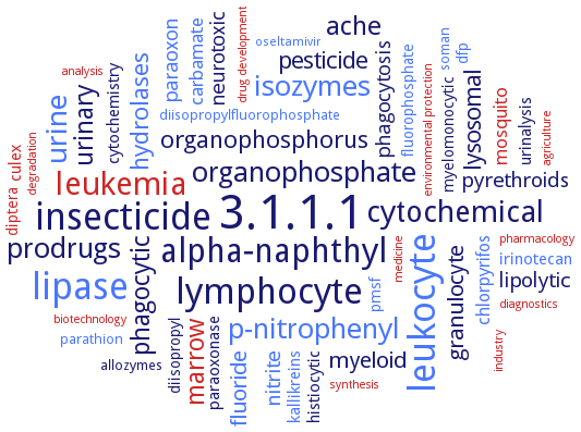
Word Map on EC 3.1.1.1 
-
3.1.1.1
-
lipase
-
insecticide
-
leukocyte
-
lymphocyte
-
alpha-naphthyl
-
leukemia
-
cytochemical
-
urine
-
isozymes
-
p-nitrophenyl
-
organophosphate
-
prodrugs
-
ache
-
phagocytic
-
marrow
-
urinary
-
organophosphorus
-
hydrolases
-
pesticide
-
lysosomal
-
fluoride
-
myeloid
-
granulocyte
-
lipolytic
-
pyrethroids
-
nitrite
-
neurotoxic
-
paraoxon
-
phagocytosis
-
carbamate
-
mosquito
-
chlorpyrifos
-
paraoxonase
-
histiocytic
-
urinalysis
-
cytochemistry
-
kallikreins
-
culex
-
irinotecan
-
dfp
-
fluorophosphate
-
diptera
-
diisopropyl
-
myelomonocytic
-
pmsf
-
soman
-
parathion
-
allozymes
-
diisopropylfluorophosphate
-
oseltamivir
-
industry
-
environmental protection
-
agriculture
-
analysis
-
diagnostics
-
synthesis
-
degradation
-
biotechnology
-
medicine
-
drug development
-
pharmacology
- 3.1.1.1
- lipase
-
insecticide
- leukocyte
- lymphocyte
-
alpha-naphthyl
- leukemia
-
cytochemical
- urine
- isozymes
- p-nitrophenyl
-
organophosphate
-
prodrugs
-
ache
- phagocytic
- marrow
- urinary
-
organophosphorus
- hydrolases
-
pesticide
- lysosomal
- fluoride
- myeloid
- granulocyte
-
lipolytic
-
pyrethroids
- nitrite
-
neurotoxic
- paraoxon
-
phagocytosis
- carbamate
- mosquito
- chlorpyrifos
- paraoxonase
- histiocytic
-
urinalysis
-
cytochemistry
- kallikreins
- culex
- irinotecan
- dfp
- fluorophosphate
- diptera
-
diisopropyl
-
myelomonocytic
- pmsf
- soman
- parathion
-
allozymes
- diisopropylfluorophosphate
- oseltamivir
- industry
- environmental protection
- agriculture
- analysis
- diagnostics
- synthesis
- degradation
- biotechnology
- medicine
- drug development
- pharmacology
Reaction
Synonyms
4-nitrophenyl esterase, ACAT, Acetyl esterase, acetyl xylan esterase, acid retinyl ester hydrolase, AcXE, Acyl coenzyme A:cholesterol acyltransferase, acylcarnitine hydrolase, AF1716, AF1763, AFEST, ali-esterase, aliesterase, alpha-carboxylesterase, alpha-esterase 7, alphaE7, alphaEsterase7, AOPP herbicide carboxylesterase, APE1547, AREH, aspirin hydrolase, Axe1NaM1, Axe1NaM2, Axe1NaM3, B-esterase, Bacillus esterase, BdB1, BioHe, BioHs, Brain carboxylesterase hBr1, BSE, BsE-NP01, BSE01701, butyrate esterase, butyryl esterase, CaE, CaesCCR11, carboxyesterase, Carboxyesterase ES-10, carboxyl ester hydrolase, carboxyl esterase, carboxyl/cholinesterase, carboxylate esterase, carboxylesterase, carboxylesterase 1, carboxylesterase 1A2, carboxylesterase 1C, carboxylesterase 2, carboxylesterase B, carboxylesterase B1, carboxylesterase-1, carboxylesterase-2, Carboxylesterase-5C, carboxylic acid esterase, carboxylic ester hydrolase, carboxylic esterase, Carboxylic-ester hydrolase, CarE, CbE, CCE, CE, CE-1, CE-2, CE1, CE2, CE21p, CE7 AcXE, CEH, CES, CES 1C, CES-2, CES1, CES1-b, CES1-c, CES1A, CES1A1, CES1A2, Ces1c, Ces1d, Ces1e, Ces1g, CES2, CES2A1, Ces3-1, Ces3a, cholesteryl ester hydrolase, cocaine esterase, cocaine:benzoylecgonine esterase, COE, CPT-CE, CXE, CXE1, CXE10, D1CarE5, de-arenethiolase, DEHP, E3 carboxylesterase, E34Tt, EC 3.1.1.21, Egasyn, ES-10, Es-22, ES-HTEL, ES-HVEL, ES-Male, ES-THET, ES10, ES11, ES3, ES4, ES5, ES6, ES7, ES9, EST, EST-3, EST-4, EST-5A, EST-5B, EST-5C, Est-AF, Est0071, Est1, Est1C, EST2, ESt3, Est3 esterase, Est30, Est55, EstA, EstA esterase, EstB, estBB1, EstC, EstC1, esterase, esterase 1F, esterase 2, esterase 2B, esterase 6A, esterase 9A, esterase A, esterase B, esterase B1, esterase D, esterase Sso2518, esterase, carboxyl, Esterase-22, Esterase-31, esterase-6, EStFa, EstGtA2, EstP, EstPS1, EstSt7, EstU1, Fluazifop-P-butyl carboxylesterase, Fluazifop-P-butyl hydrolase, FpbH, fungus-inducible pepper carboxylesterase, general odorant degradation enzyme, GODE, hCE-2, HCE1, HCE2, hCES1, hiCE, HMSE, hormone-sensitive lipase, HSL, human carboxylesterase 1, human carboxylesterase 2, human liver carboxylesterase, ICE, ICXE14, JHE-related, JHER, kidney bean esterase, Kidney microsomal carboxylesterase, LcalphaE7, LIP4, LipA, LipC, Liver microsomal carboxylesterase, malathion carboxylesterase, MAP esterase, MCE, ME, MeIAA esterase, MeSA esterase, methylbutyrase, methylbutyrate esterase, mfCES2v3, Microsomal palmitoyl-CoA hydrolase, monobutyrase, Monocyte/macrophage serine esterase, More, mouse carboxylesterase, MT2282, NaM1, NaM2, NaM3, Non-specific carboxylesterase, nonspecific carboxylesterase, ODE, odorant degradation enzyme, P186_1588, PA3859, PepEST, PF2001, Pf2001 esterase, PI 5.5 esterase, PI 6.1 esterase, pig liver esterase, PLE, PMPMEase, pnbA, PnbA1, PnbA2, polyisoprenylated methylated protein methyl esterase, porcine liver carboxylesterase, PPE, PPMTase, PrbA, prenylated methylated protein methyl esterase, procaine esterase, Proline-beta-naphthylamidase, propionyl esterase, pyrethroid-hydrolysing esterase, Rsp3690, SABP2's methyl salicylate esterase, salicylic acid-binding protein 2, SeE, serine protease-like, serine-type carboxylesterase, SexiCXE10, SiRe0290, SshEstI, SshEstI esterase, Sso P1 carboxylesterase, Sso-EST1, SSO2493, SSO2517, SSoP1 CE, SsoPEst, ST0071, ST2026, STK_00710, Sto-Est, TGH, triacetin esterase, triacylglycerol hydrolase, vitamin A esterase, VmoLac


 results (
 results (
 top
top


