2.7.1.40: pyruvate kinase
This is an abbreviated version!
For detailed information about pyruvate kinase, go to the full flat file.
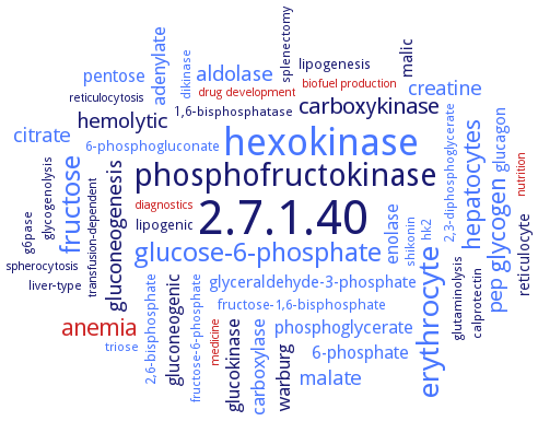
Word Map on EC 2.7.1.40
-
2.7.1.40
-
hexokinase
-
phosphofructokinase
-
erythrocyte
-
fructose
-
glucose-6-phosphate
-
hepatocytes
-
glycogen
-
anemia
-
carboxykinase
-
gluconeogenesis
-
aldolase
-
pep
-
hemolytic
-
malate
-
citrate
-
creatine
-
glucokinase
-
adenylate
-
carboxylase
-
warburg
-
gluconeogenic
-
enolase
-
phosphoglycerate
-
pentose
-
6-phosphate
-
malic
-
glucagon
-
glyceraldehyde-3-phosphate
-
reticulocyte
-
lipogenesis
-
6-phosphogluconate
-
fructose-1,6-bisphosphate
-
lipogenic
-
2,6-bisphosphate
-
g6pase
-
1,6-bisphosphatase
-
liver-type
-
splenectomy
-
hk2
-
dikinase
-
glutaminolysis
-
calprotectin
-
triose
-
shikonin
-
2,3-diphosphoglycerate
-
fructose-6-phosphate
-
glycogenolysis
-
drug development
-
medicine
-
biofuel production
-
reticulocytosis
-
transfusion-dependent
-
spherocytosis
-
nutrition
-
diagnostics
- 2.7.1.40
- hexokinase
-
phosphofructokinase
- erythrocyte
- fructose
- glucose-6-phosphate
- hepatocytes
- glycogen
- anemia
-
carboxykinase
-
gluconeogenesis
- aldolase
- pep
-
hemolytic
- malate
- citrate
- creatine
- glucokinase
- adenylate
- carboxylase
-
warburg
-
gluconeogenic
- enolase
- phosphoglycerate
- pentose
- 6-phosphate
-
malic
- glucagon
- glyceraldehyde-3-phosphate
- reticulocyte
-
lipogenesis
- 6-phosphogluconate
- fructose-1,6-bisphosphate
-
lipogenic
- 2,6-bisphosphate
- g6pase
-
1,6-bisphosphatase
-
liver-type
-
splenectomy
- hk2
- dikinase
-
glutaminolysis
-
calprotectin
- triose
- shikonin
- 2,3-diphosphoglycerate
- fructose-6-phosphate
-
glycogenolysis
- drug development
- medicine
- biofuel production
-
reticulocytosis
-
transfusion-dependent
-
spherocytosis
- nutrition
- diagnostics
Reaction
Synonyms
ATP/pyruvate O'-phosphotransferase, ATP:pyruvate 2-O-phosphotransferase, CPK, CPK1, cPK2, cPK3, cPK4, cPK5, CTHBP, cytosolic pyruvate kinase, cytosolic thyroid hormone binding protein, EHI_098420, EhPK, EhPyk, erythroid (R-type) pyruvate kinase, fluorokinase, hL-PYK, hLPYK, hPKM2, K+-dependent PK, K+-independent PK, kinase, fluoro- (phosphorylating), kinase, pyruvate (phosphorylating), L-PK
ECTree

Advanced search results
Crystallization
Crystallization on EC 2.7.1.40 - pyruvate kinase
Please wait a moment until all data is loaded. This message will disappear when all data is loaded.
 top
topCRYSTALLIZATION (Commentary) 

ORGANISM
UNIPROT
LITERATURE
purified recombinant untagged enzyme in apoform or complexed with the non-hydrolyzable ATP analogue adenyl-5'-yl imidodiphosphate, hanging drop vapor diffusion technique, 4°C, mixing of 7 mg/ml protein in 0.65 M ammonium sulfate and 0.1 M sodium acetate, pH 4.0, and with 3 mM adenyl-5'-yl imidodiphosphate, 5 mM magnesium chloride, and 5 mM pyruvic acid for the enzyme complex, X-ray diffraction structure determination and analysis at 2.5 A resolution, molecular replacement
mutant C9S/C268S, 2.4 A resolution. Crystal belongs to space group P6222. Enzyme is a homotetramer, a sulfate ion is bound in a pocket in the effector-binding C domain
purified enzyme mutant S12D as S12D-L-PYK-Fru-1,6-bisphosphate-Mn-Na-citrate complex, vapor diffusion hanging drop method, PYK S12D at 4 mg/ml in 10 mM MES, pH 6.8, 5 mM MgCl2, 10 mM KCl, and 2 mM DTT, 52 mM Na-ATP, and 1.3 mM Na-Fru-1,6-BP, mixing with well solution in varied ratios of 2:2, 4:2, 2:4, and 4:4, the well solution contains 50 mM sodium citrate, pH 4.9, 26 mM MnCl2, and 3-5% PEG 6000, X-ray diffraction structure determination and analysis at 1.8-1.95 A resolution, molecular replacement and modeling
purified recombinant N-terminally His6-tagged mutant enzymes, hanging drop vapour diffusion method, mixing of 0.0015 ml of 2.0-6.0 mg/ml protein in 50 mM MES, pH 6.8, 100 mM KCl, 10% glycerol, and 2 mM DTT, with 0.0015 ml of reservoir solution containing 200 mM ammonium citrate dibasic, and 20% PEG 3350 for mutant D499N, or 325 mM ammonium citrate dibasic, and 16% PEG 3350 for mutant W527H, or 200 mM ammonium citrate, pH 6.0, and 24% PEG 3350 for mutant DELTA529/S531G, or 200 mM ammonium citrate, pH 5.6, and 16% PEG 3350 for mutant S531E, and equilibration against 1 ml of reservoir solution, at 25°C, X-ray diffraction structrue determination and analysis at 2.15-2.42 A resolution, molecular replacement using the A and C domains of chain A of the S12D variant of hLPYK (PDB ID 4ip7) as a rigid-body model, modeling
purified recombinant PHD3/PKM2 complex, hanging drop vapor diffusion method, mixing of 5 mg/ml protein in 20 mM MES, pH 6.0, 75 mM NaCl, and 1 mM DTT, in a 1:1 ration with reservoir solution, 18°C, two weeks, X-ray diffraction structure determination and analysis at 3.0 A resolution, molecular replacement method and modeling
the X-ray structure of human hPKM2 complexed with Mg2+, K+, the inhibitor oxalate, and the allosteric activator D-fructose 1,6-bisphosphate determined to a resolution of 2.82 A. (His)6-tagged hPKM2 complex, hanging drop vapor diffusion
crystals grown with ammonium sulfate as precipitant adopt an active-like conformation, with sulfate ions at the active and effector sites. Crystal soaking in sulfate-free buffers induces major conformational changes in the tetramer. The unwinding of the Aalpha6' helix and the inward hinge movement of the B domain are coupled with a significant widening of the tetramer caused by lateral movement of the C-domains
enzyme complexed with inhibitor 4-[(1,1-dioxo-1,2-benzothiazol-3-yl)sulfanyl]benzoic acid, mxing of 0.0015 ml of 10 mg/ml protein in 20 mM TEA, pH 7.2, 1 mM 1,3,6,8-pyrenetetrasulfonic acid, and 9 mM inhibitor, with 0.0015 ml of well solution containing 12-16% PEG 8000, 20 mM TEA buffer, pH 7.2, 50 mM magnesium chloride, 100 mM potassium chloride, and 10% glycerol, 1-2 days, and equilibration for 14 h over a well solution composed of 14-18% PEG 8,000, 20 mM TEA buffer, pH 7.2, 50 mM magnesium chloride, 100 mM potassium chloride, and 25% glycerol, X-ray diffraction structure determination and analysis at 2.65 A resolution
-
purified recombinant wild-type and mutant enzymes free and in complex with ligands ATP, oxalate, and fructose 2,6-bisphosphate, hanging drop vapour diffusion method, 0.0015 ml of 15 mg/ml protein in 20 mM TEA, pH 7.2, are mixed with 0.0015 ml of well solution composed of 10-16% PEG 8000, 20 mM TEA, pH 7.2, 50 mM MgCl2, 100 mM KCl, and 10-15% glycerol, 4°C or 17°C, 1week, X-ray diffraction structure determination and analysis
hanging drop vapour diffusion method using 60 mM succinate (pH 5.5), 5.8 mM sodium pyruvate, 2.4 mM MnCl2, 450 mM KCl, and a range of 18 to 20% PEG 8000
purified isozyme PYK-I in complex with substrate analogue oxalate, activator glucose 6-phosphate, and product ATP, hanging drop vapour diffusion method, mixing of 0.001 ml of protein solution and 500 nl of 20 mM ligand solution, with 0.0015 ml of reservoir solution containing 12% PEG 8000, 10-20% glycerol, 50 mM TEA, pH 7.2, 100 mM KCl, and 50 mM MgCl2, and equilibration against 1 ml of reservoir solution, X-ray diffraction structure determination and analysis, molecular replacement using the structure obtained from the deposited T-state PfPYK (PDB ID 3KHD) as template, molecular modelling
hanging-drop vapor diffusion method at 18°C, crystal structure is determined to 2.2 A resolution
liver L-type isozyme, erythrocytic R-type isozyme, M1- and M2-type isozymes
-
purified recombinant N-terminally His6-tagged full-length and N-terminaly truncated enzymes, with the B domain in the open and closed conformations, crystals of the truncated TgPK1 are grown in the presence of K+, Mg2+, and inhibitor, X-ray diffraction structure determination and analysis
structure analysis of TbrPYK tetramer complexed with fructose 2,6-bisphosphate (PDB ID 4HYV) and comparison with the tetrameric structure of TcoPYK (PDB IDs 6SU1 and 6SU2)
purified recombinant His-tagged enzyme free and in complex with fructose 1,6-bisphosphate, hanging drop vapor diffusion method, mixing of 0.002 ml of 4 mg/ml protein solution with 0.002 ml of reservoir solution containing 100 mM ammonium citrate dibasic, and 12.5% PEG 3350, and equilibration against 0.150 ml reservoir solution, 20°C, method optimization, X-ray diffraction structure deterination and analysis, molecular replacement using the structure of TbrPYK tetramer complexed with fructose 2,6-bisphosphate (PDB ID 4HYV) as a search model, modeling
purified recombinant untagged enzyme, synchrotron radiation small-angle X-ray scattering structure determination and analysis
-