Any feedback?
Please rate this page
(all_enzymes.php)
(0/150)

BRENDA support

2.3.1.179: beta-ketoacyl-[acyl-carrier-protein] synthase II

This is an abbreviated version!
For detailed information about beta-ketoacyl-[acyl-carrier-protein] synthase II, go to the full flat file.
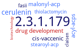
Word Map on EC 2.3.1.179

Reaction

a (Z)-hexadec-9-enoyl-[acyl-carrier protein]
+
a malonyl-[acyl-carrier protein]
=
a (Z)-3-oxooctadec-11-enoyl-[acyl-carrier protein]
+
CO2
+
an [acyl-carrier protein]

Synonyms

3-ketoacyl acyl synthase II, 3-ketoacyl-ACP synthase, 3-ketoacyl-ACP synthase 2, 3-ketoacyl-ACP synthase II, 3-oxoacyl-(acylcarrier protein) synthase II, 3-oxoacyl-ACP synthase II, B-ketoacyl-ACP synthase II, beta-ketoacyl ACP-synthase II, beta-ketoacyl acyl carrier protein synthase II, beta-ketoacyl acyl-carrier protein synthase II, beta-ketoacyl synthase II, beta-ketoacyl-(acyl-carrier-protein) synthase II, beta-ketoacyl-ACP synthase FabF3, beta-ketoacyl-ACP synthase II, beta-ketoacyl-acyl carrier protein synthase I/II, beta-ketoacyl-acyl carrier protein synthase II, beta-ketoacyl-acyl carrier protein synthases II, beta-ketoacyl-acyl carrier protein synthetase II, beta-ketoacyl-acyl-carrier protein synthase II, beta-ketoacyl-acyl-carrier-protein synthase II, beta-ketoacyl-[ACP] synthase II, beta-ketoacyl-[ACP] synthase-II, beta-ketoacyl-[acyl carrier protein (ACP)] synthase II, beta-ketoacyl-[acyl-carrier protein (ACP)] synthase II, beta-ketoacyl-[acyl-carrier-protein] synthase II, FabB, FabB/F, FabF, FabF elongation condensing enzyme, FabF of type II fatty acid biosynthesis, FabF1, FASII, fatty acid synthesis type II, KAS II, KAS-II, KAS2, KASII

ECTree

     2 Transferases
         2.3 Acyltransferases
             2.3.1 Transferring groups other than aminoacyl groups
                2.3.1.179 beta-ketoacyl-[acyl-carrier-protein] synthase II

Crystallization

Crystallization on EC 2.3.1.179 - beta-ketoacyl-[acyl-carrier-protein] synthase II

Please wait a moment until all data is loaded. This message will disappear when all data is loaded.
CRYSTALLIZATION (Commentary)
ORGANISM
UNIPROT
LITERATURE
crystal structure determination and analysis at 2.6 A resolution
hanging drop vapour diffusion method at room temperature, using 27% PEG 8000 as precipitant and buffered at pH 7.5 with 0.1 M HEPES. Crystal structure is determined with the multiple isomorphous replacement method and refined at 2.4 A resolution, space group P3(1)21
purified recombinant free wild-type enzyme, and mutant C164Q enzyme, free or in complex with 3-(benzoylamino)-2-hydroxybenzoic acid, mixing of 0.001 ml of 20 mg/ml protein in 25 mM Tris-HCl, pH 7.5, 150 mM NaCl, with 0.001 ml reservoir solution containing 0.2 M MgCl2, 0.1 M Tris-HCl, pH 7.0, 10% w/v PEG 8000 for the wild-type enzyme, or 0.001 ml of 20 mg/ml protein in 50 mM Na2HPO4, pH 7.8, 150 mM NaCl, 10% v/v glycerol, and 0.5 mM DTT with 0.001 ml of reservoir consisting of 0.2 M NH4HCO2, 25% w/v PEG 3350 for the mutant enzyme, equilibration against 0.06 ml reservoir solution, at 20°C, X-ray diffraction structure determination and analysis at 1.67-2.46 A resolution, molecular replacement
hanging drop vapor diffusion method. 1.3 A resolution crystal structure
-
hanging-drop vapour-diffusion method
purified recombinant mutant H303A from 20% polyethylene glycol 3350, 0.2 M potassium acetate, X-ray diffraction structure determination and analysis
-
purified recombinant wild-type and mutant E383A enzymes, hanging-drop vapour-diffusion method at room temperature, 0.001 ml of protein solution, containing 10 mg/mlprotein in 20 mM Tris-HCl, pH 8.0, 50 mM NaCl, and 10% glycerol, is mixed with 0.001 ml of precipitating solution, containing 0.2 M sodium acetate, 0.1 M Tris-HCl, pH 8.5, and 30% PEG 4000, formation of different crystal forms, X-ray diffraction structure determinations and analysis at 1.3-2.1 A resolution
sitting drop vapor diffusion method, using either 0.1 M sodium chloride, 0.1 M bicine pH 9.0, 20% (w/v) PEG MME 550 or 0.1 M HEPES pH 7.5, 12% (w/v) PEG 3350 or 0.1 M Bis-Tris pH 5.5, 25% (w/v) PEG 3350, 0.2 M ammonium sulfate
rod-shaped crystals of the purified enzyme are grown in two weeks by the hanging-drop vapor-diffusion method, 1.54 A resolution, space group P3(1)21, cells dimensions: a = b = 100.8 A, c = 74.7 A
purified recombinant enzyme, hanging drop vapour diffusion technique, mixing of 0.0015 ml of 21 mg/ml protein solution with an equal amount of reservoir solution, containing 0.2 M lithium chloride, 0.1 M HEPES sodium salt pH 7.0, and 24% PEG 6000, and equilibration against 0.3 ml of reservoir solution, 13°C, X-ray diffraction structure determination and analysis at 2.7 A resolution, molecular replacement using the enzyme structure of Escherichia coli, PDB ID 1KAS, as search model
Q7CJ22