1.8.98.2: sulfiredoxin
This is an abbreviated version!
For detailed information about sulfiredoxin, go to the full flat file.

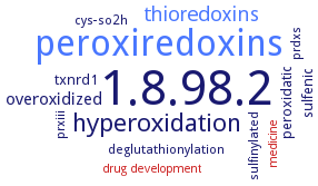
Word Map on EC 1.8.98.2 
-
1.8.98.2
-
peroxiredoxins
-
hyperoxidation
-
thioredoxins
-
overoxidized
-
sulfenic
-
peroxidatic
-
txnrd1
-
sulfinylated
-
deglutathionylation
-
prxiii
-
prdxs
-
cys-so2h
-
medicine
-
drug development
- 1.8.98.2
- peroxiredoxins
-
hyperoxidation
- thioredoxins
-
overoxidized
-
sulfenic
-
peroxidatic
- txnrd1
-
sulfinylated
-
deglutathionylation
-
prxiii
-
prdxs
-
cys-so2h
- medicine
- drug development
Reaction
Synonyms
AtSrx, cysteine-sulfinic acid reductase, neoplastic progression 3, peroxiredoxin-(S-hydroxy-S-oxocysteine) reductase, protein cysteine sulfinic acid reductase, Srx, Srx1, Srxn1, sulfiredoxin, sulfiredoxin 1, sulfiredoxin-1, sulphiredoxin
ECTree
Advanced search results
Crystallization
Crystallization on EC 1.8.98.2 - sulfiredoxin
Please wait a moment until all data is loaded. This message will disappear when all data is loaded.
purified enzyme AtSrx in complex with ADP, sitting drop vapor diffusion method, mixing of 0.001 ml of 20 mg/ml protein in 50 mM Tris-HCl, pH 7.5, 50 mM NaCl, and 1 mM DTT, with 0.001 ml of well solution containing 0.8 M NaH2PO4/1.2M KH2PO4, and acetate, pH 4.5, at 16°C, 1 week, X-ray diffraction structure determination and analysis at 3.20 A resolution
crystals of the wild-type and SeMet forms of ET-hSrx were obtained by the vapor diffusion method. 1.65 A crystal structure of human Srx
purified Srx-Prx1 complex, hanging drop vapor diffusion method, mixing of 10 mg/ml protein in 20 mM HEPES, pH 7.5, 100 mM NaCl with well solution containing 100 mM citric acid, pH 4.5, 26% PEG 400, and 100 mM CsCl, X-ray diffraction structure determination and analysis at 3.0 A resolution, molecular replacement and modeling by superimposing five dimeric Srx-Prx1 complexes (PDB entry 2RII) onto the five Prx dimers within the decameric Prx2-SO2H (PDB ID 1QMV) structure. The entire active site helix of Prx1 (residues 46-69) and the C-terminus (residues 169-199) are removed from the search model to reduce bias. Two decamers, each containing ten Prx and ten Srx molecules (five Prx dimers and ten Srx monomers), are found in the asymmetric unit, consistent with the observed self-rotation function. Additional crystallization of hyperoxidized Prx2 and Prx3 variants
vapour diffusion method, 2.6 A crystal structure of the sulphiredoxinperoxiredoxin-I complex


 results (
 results (
 top
top


