1.8.1.7: glutathione-disulfide reductase
This is an abbreviated version!
For detailed information about glutathione-disulfide reductase, go to the full flat file.

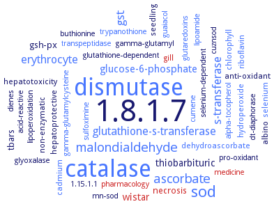
Word Map on EC 1.8.1.7 
-
1.8.1.7
-
dismutase
-
catalase
-
sod
-
malondialdehyde
-
ascorbate
-
s-transferase
-
gst
-
glutathione-s-transferase
-
erythrocyte
-
glucose-6-phosphate
-
wistar
-
thiobarbituric
-
gsh-px
-
tbars
-
necrosis
-
selenium
-
dehydroascorbate
-
anti-oxidant
-
hydroperoxide
-
cadmium
-
albino
-
hepatoprotective
-
hepatotoxicity
-
riboflavin
-
non-enzymatic
-
seedling
-
gill
-
chlorophyll
-
dt-diaphorase
-
cumene
-
medicine
-
selenium-dependent
-
lipoperoxidation
-
sulfoximine
-
glyoxalase
-
gamma-glutamyl
-
alpha-tocopherol
-
transpeptidase
-
pro-oxidant
-
cuznsod
-
dienes
-
trypanothione
-
glutaredoxins
-
glutathione-dependent
-
guaiacol
-
buthionine
-
1.15.1.1
-
acid-reactive
-
mn-sod
-
pharmacology
-
lipoamide
-
gamma-glutamylcysteine
- 1.8.1.7
- dismutase
- catalase
- sod
- malondialdehyde
- ascorbate
- s-transferase
- gst
- glutathione-s-transferase
- erythrocyte
- glucose-6-phosphate
- wistar
-
thiobarbituric
- gsh-px
-
tbars
- necrosis
- selenium
- dehydroascorbate
-
anti-oxidant
- hydroperoxide
- cadmium
-
albino
-
hepatoprotective
-
hepatotoxicity
- riboflavin
-
non-enzymatic
- seedling
- gill
- chlorophyll
- dt-diaphorase
- cumene
- medicine
-
selenium-dependent
-
lipoperoxidation
- sulfoximine
- glyoxalase
-
gamma-glutamyl
- alpha-tocopherol
- transpeptidase
-
pro-oxidant
-
cuznsod
-
dienes
- trypanothione
- glutaredoxins
-
glutathione-dependent
- guaiacol
-
buthionine
-
1.15.1.1
-
acid-reactive
- mn-sod
- pharmacology
- lipoamide
- gamma-glutamylcysteine
Reaction
2 glutathione
+
Synonyms
At3g54660, EC 1.6.4.2, GLR, glutahione reductase, glutathione disulfide reductase, glutathione reductase, glutathione reductase (NADPH), glutathione reductase 3, glutathione reductase Glr1, glutathione S-reductase, glutathione: NADP(+) oxidoreductase, glutathione: NADP+ oxidoreductase, glutathione:NADP+ oxidoreductase, Gor, GOR1, GOR2, GR, GR1, GR2, Gr3, GRase, GRase-1, GRd, GSH reductase, GSHR, Gsr, GSR-1, GSSG reductase, HCOI_01258400, hGR, HvGR1, HvGR2, multifunctional thioredoxin-glutathione reductase, NADPH-glutathione reductase, NADPH-GSSG reductase, NADPH-reduced GR, NADPH: oxidized glutathione oxidoreductase, NADPH:oxidized glutathione oxidoreductase, NADPH:oxidized-glutathione oxidoreductase, OBP29, PfGR, psgr, PtGR1.1, PtGR1.2, PtGR2, reductase, glutathione, SpGR, TaGR1, TaGR2, TGR, thioredoxin glutathione reductase, thioredoxin/glutathione reductase, TrxR3


 results (
 results (
 top
top


