1.3.5.1: succinate dehydrogenase
This is an abbreviated version!
For detailed information about succinate dehydrogenase, go to the full flat file.
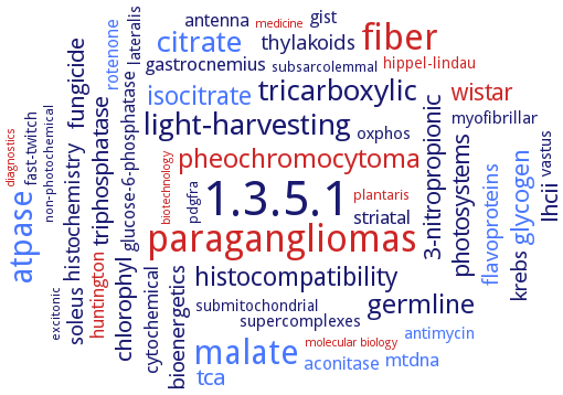
Word Map on EC 1.3.5.1
-
1.3.5.1
-
paragangliomas
-
fiber
-
malate
-
atpase
-
citrate
-
light-harvesting
-
tricarboxylic
-
pheochromocytoma
-
germline
-
histocompatibility
-
isocitrate
-
glycogen
-
3-nitropropionic
-
photosystems
-
wistar
-
lhcii
-
fungicide
-
chlorophyl
-
tca
-
triphosphatase
-
krebs
-
flavoproteins
-
histochemistry
-
soleus
-
bioenergetics
-
thylakoids
-
huntington
-
striatal
-
gastrocnemius
-
cytochemical
-
mtdna
-
aconitase
-
glucose-6-phosphatase
-
antenna
-
rotenone
-
gist
-
antimycin
-
oxphos
-
lateralis
-
vastus
-
myofibrillar
-
supercomplexes
-
submitochondrial
-
hippel-lindau
-
fast-twitch
-
pdgfra
-
subsarcolemmal
-
plantaris
-
non-photochemical
-
excitonic
-
biotechnology
-
medicine
-
diagnostics
-
molecular biology
- 1.3.5.1
- paragangliomas
- fiber
- malate
- atpase
- citrate
-
light-harvesting
-
tricarboxylic
- pheochromocytoma
-
germline
-
histocompatibility
- isocitrate
- glycogen
-
3-nitropropionic
- photosystems
- wistar
- lhcii
-
fungicide
-
chlorophyl
- tca
- triphosphatase
-
krebs
- flavoproteins
-
histochemistry
- soleus
-
bioenergetics
- thylakoids
- huntington
- striatal
- gastrocnemius
-
cytochemical
- mtdna
- aconitase
- glucose-6-phosphatase
- antenna
- rotenone
-
gist
- antimycin
-
oxphos
- lateralis
-
vastus
- myofibrillar
-
supercomplexes
-
submitochondrial
- hippel-lindau
-
fast-twitch
-
pdgfra
-
subsarcolemmal
- plantaris
-
non-photochemical
-
excitonic
- biotechnology
- medicine
- diagnostics
- molecular biology
Reaction
Synonyms
8-methylmenaquinol:fumarate reductase, AaSdhB, bacterial succinate:quinone oxidoreductase flavoprotein, complex II, Complex II homolog, complex II of the respiratory chain, complex II succinate:ubiquinone oxidoreductase, DCPIP oxidoreductase, dehydrogenase, succinate, dehydrogenase/complex II, EC 1.3.5.4, EC 1.3.99.1, Fcc3, fdrB, FL cyt, Flavocytochrome c3, FRD, FrdA, FRdABCD, FrdC, FRdCAB, FrdD, fumarate reductase, fumarate reductase complex, fumaric hydrogenase, Ifc3, Iron(III)-induced flavocytochrome C3, iron-sulfur subunit of succinate dehydrogenase, menaquinol-1 fumarate reductase, menaquinol-fumarate oxidoreductase, menaquinol:fumarate oxidoreductase, methylmenaquinol:fumarate reductase, MFR, MFR complex, MfrA, MfrB, mitochondrial complex II, mitochondrial succinate dehydrogenase, mitochondrial succinate:ubiquinone oxidoreductase, More, mQFR, MSMEG_0416, MSMEG_0417, MSMEG_0418, MSMEG_0419, MSMEG_0420, MSMEG_1669, MSMEG_1670, MSMEG_1671, MSMEG_1672, non-classical succinate:quinone reductase, QFR, quinol-fumarate reductase, quinol:fumarate reductase, SDG, SDG-1, SDG-2, SDH, SDH1, SDH2, SDH2-1, SDH2-2, SDH3, SDH4, SdhA, sdhABE, SDHB, SdhC, sdhCAB, SdhCDAB, SdhD, SDISP, SQR, succinate dehydrogenase, succinate dehydrogenase (caldariellaquinone), succinate dehydrogenase (quinone), succinate dehydrogenase B, succinate dehydrogenase complex, succinate dehydrogenase flavoprotein subunit Sdh1p, succinate dehydrogenase iron-sulfur protein, succinate dehydrogenase iron-sulphur protein, succinate dehydrogenase subunit B, succinate oxidoreductase, succinate-2,6-dichlorophenolindophenol oxidoreductase, succinate-coenzyme Q reductase, succinate-quinone oxidoreductase, succinate-quinone reductase, succinate-ubiquinone oxidoreductase, succinate:caldariellaquinone oxidoreductase, succinate:menaquinone 7-reductase, succinate:menaquinone oxidoreductase, succinate:menaquinone reductase, succinate:MK reductase, succinate:quinone oxidoreductase, succinate:quinone reductase, succinate:quinone reductases, succinate:ubiquinone oxidoreductase, succinate:ubiquinone reductase, succinic acid dehydrogenase, succinic dehydrogenase, succinodehydrogenase, succinyl dehydrogenase, Tneu_0423
ECTree

Advanced search results
Cofactor
Cofactor on EC 1.3.5.1 - succinate dehydrogenase
Please wait a moment until all data is loaded. This message will disappear when all data is loaded.
 top
topCOFACTOR 

ORGANISM
UNIPROT
COMMENTARY 
LITERATURE
IMAGE
heme b556
-
ubiquinone reduction by an electron transfer relay comprising a flavin adenine dinucleotide cofactor, three iron-sulfur clusters, and possibly a heme b556. At the heart of the electron transport chain is a [4Fe-4S] cluster with a low midpoint potential that acts as an energy barrier against electron transfer
heme b562
-
the enzyme has a heme b-containing membrane-anchoring dimer, comprising the Sdh3p and Sdh4p subunits, overview
menaquinone
Megalodesulfovibrio gigas
one menaquinone molecule is bound near heme bL in the hydrophobic subunit C
quinone
-
with a periplasmically oriented quinone binding site of the enzyme
cytochrome b
-
cytochrome b-558, 3.6 nmol per mg of protein, functions as electron carrier between NADH dehydrogenase and succinate dehydrogenase in the Ascaris NADH-fumarate reductase system
-
cytochrome b
-
cytochrome b-558, is the smallest protein of the complex with MW: 19000, it is a transmembrane protein and anchors succinate dehydrogenase to the cytoplasmic side of the membrane
-
cytochrome b
-
has a MW of 25000 Da, a midpoint potential of - 15 mV and is reducible by dimethylnaphthohydroquinone in the absence of the other subunits
-
cytochrome b
-
the enzyme contains a cytochrome b with a midpoint potential of -20 mV, referred to as the high-potential cytochrome b and a cytochrome b with a midpoint potential of -200 mV, referred to as the low-potential cytochrome b
-
cytochrome b
-
the isolated two-subunit membrane anchoring protein contains 35 nmol cytochrome b-556 per mg protein
-
cytochrome b
-
cytochrome b-558, composed of two hydrophobic polypeptides with molecular masses of 17.2 and 12.5 kDa, which correspond to the two small subunits of complex II
-
FAD
-
ubiquinone reduction by an electron transfer relay comprising a flavin adenine dinucleotide cofactor, three iron-sulfur clusters, and possibly a heme b556. At the heart of the electron transport chain is a [4Fe-4S] cluster with a low midpoint potential that acts as an energy barrier against electron transfer
FAD
-
flavoprotein, 41 pmol FAD per mg of protein in wild-type strain YPH499, possesses a flavoprotein subunit Sdh1p as part of the catalytic dimer of the tetrameric enzyme
FAD
-
FAD is non-covalently attached to SdhA. The reason for the lack of succinate oxidation activity might be explained by the absence of a covalently bound FAD which seems to be a prerequisite for succinate oxidation activity
FAD
-
proteins displays two redox active domains, one containing four c-type hemes (I-IV) and another containing FAD at the catalytic site. Redox titrations followed by NMR and visible spectroscopies are applied to investigate the properties that allow a chain of single-electron co-factors to sustain the activity of a multielectron catalytic site. The results show that the redox behaviour of fumarate reductases is dominated by a strong interaction between hemes II and III. This interaction facilitates a sequential transfer of two electrons from the heme domain to FAD via heme IV
FAD
-
non-covalenly bound. In the enzyme containing a mutant A86H flavoprotein subunit the FAD is covalently bound
FAD
-
prosthetic group of fumarate reductase is covalently bound FAD. The specific activity of fumarate reductase is increased to the same extent as the content of the covalently bound FAD when the membrane is fractionated with cholate and ammonium sulfate. The acid-extractable FAD is removed by this procedure
FAD
-
stoichiometric ratio between covalently bound FAD and the iron-sulfur cluster is 1:1. Protoheme IX is present in about 2:1 stoichiometry to covalently bound FAD
FAD
-
FAD of Sdh1 is covalently attached at an active site His residue, 8alpha-N3-histidyl-FAD linkage, involving Arg582, required for enzyme activity. Flavinylation and assembly of succinate dehydrogenase are dependent on the C-terminal tail of the flavoprotein subunit. FAD binding is important to stabilize the Sdh1 conformation enabling association with Sdh2 and the membrane anchor subunits
FAD
-
flavinylation of SDH is dependent on SDH1:SDHA F2 interaction requiring the C-terminal tail of SDH1
FAD
the enzyme complex contains one molecule of covalently bound FAD
FAD
the FAD prosthetic group is held in the FAD binding domain by a covalent bond to His A79 and by hydrogen bonds with highly conserved residues, overview
FAD
bound to the binding site for FAD in the FAD-binding domain (residues 1-245 and 351-431), which includes a Rossmann-type fold
FAD
covalently attached FAD within the FrdA subunit, the SdhE assembly factor enhances covalent flavinylation of complex II homologues. Analysis of the mechanisms of covalent flavinylation of FrdA subunit, and propose of chemical mechanism of covalent flavinylation via a quinone-methide intermediate, overview. In Escherichia coli QFR, the flavin is covalently attached to the FrdA subunit via an 8x02 linkage to the N-epsilon of His44. This places the flavin isoalloxazine group at the interface of two domains, termed the flavin-binding domain and the capping domain. These domains can move with respect to each other, which may allow the catalytic subunit to close over the substrate-bound active site and protect the reaction intermediates from solvent. Quantitation of covalent flavinylation under aerobic and anaerobic growth conditions
FAD
covalently bound in the SDHA subunit, for electrons travel from FAD in SDHA, via three Fe-S clusters in SDHB, to the quinone-binding site at the membrane interface. Significance of FAD covalent bond versus non-covalent bond. FAD is mandatory for enzyme activity. In yeast, a soluble, mitochondrial matrix protein named Sdh5 is required for the activation and flavination of Sdh1 (SDH flavoprotein subunit homologue) as well as for SDH-dependent respiration
FAD
covalently bound in the SDHA subunit, for electrons travel from FAD in SDHA, via three Fe-S clusters in SDHB, to the quinone-binding site at the membrane interface. Significance of FAD covalent bond versus non-covalent bond. FAD is mandatory for enzyme activity. Under aerobic conditions, the FAD moiety catalyzes succinate oxidation, upon which FAD is itself reduced to FADH2. Changes in redox potential prompt electrons to move from the reduced FADH2 through Fe-S clusters in SdhB to eventually reduce ubiquinone at the ubiquinone-binding site formed by SdhC and SdhD
Fe-S center

-
the Fe-S centers in Sdh2 consist of a 2Fe-2S center proximal to the FAD site, an adjacent 4Fe-4S center followed by a 3Fe-4S center
Fe-S cluster
SDHB is an iron-sulfur cluster protein containing three Fe-S clusters
Fe-S cluster
SDHB is an iron-sulfur cluster protein containing three Fe-S clusters
Fe-S cluster
the SDHB subunit of the enzyme complex contains three iron-sulfur clusters (ISCs): [2Fe-2S], [4Fe-4S], and [3Fe-4S]
Fe-S cluster
the SDHB subunit of the enzyme complex contains three iron-sulfur clusters (ISCs): [2Fe-2S], [4Fe-4S], and [3Fe-4S]
flavin
-
quantitative determination of content in wild-type and mutant enzymes
flavin
the enzyme complex contains non-extractable flavin. The purified complex contains 4.6 nmol/mg acid non-extractable flavin
heme
-
direct role of the heme of succinate-ubiquinone oxidoreductase in transfer of electrons from the iron-sulfur cluster to the quinone
heme
-
a single heme b moiety is incorporated into the membrane anchor and only the QP-site is functional
heme
-
in a di-heme membrane anchor protein harboring two putative quinone binding sites
heme
-
proteins displays two redox active domains, one containing four c-type hemes (I-IV) and another containing FAD at the catalytic site. Redox titrations followed by NMR and visible spectroscopies are applied to investigate the properties that allow a chain of single-electron co-factors to sustain the activity of a multielectron catalytic site. The results show that the redox behaviour of fumarate reductases is dominated by a strong interaction between hemes II and III. This interaction facilitates a sequential transfer of two electrons from the heme domain to FAD via heme IV
heme
-
enzyme contains about 11 iron atoms per complex, which is expected if the enzyme contains one [2Fe-2S] cluster, one [3Fe-4S] cluster, one [4Fe-4S] cluster and two type b hemes. Protoheme IX is present in about 2:1 stoichiometry to covalently bound FAD
heme
-
enzyme contains two heme molecules. Presence of protoheme IX, absence of the other heme types. The ratio of protoheme IX to the SQR protomer is 1.5, there are two protoheme IX-binding sites in SQR
heme
-
a diheme-containing enzyme, each heterotrimer contains two heme b groups bound by the transmembrane subunit C, which are termed the proximal heme, bP, and the distal heme, bD, according to the relative proximity to the hydrophilic subunits A and B
heme
-
a diheme-containing enzyme, each heterotrimer contains two heme b groups bound by the transmembrane subunit C, which are termed the proximal heme, bP, and the distal heme, bD, according to the relative proximity to the hydrophilic subunits A and B
heme
-
a diheme-containing enzyme, each heterotrimer contains two heme b groups bound by the transmembrane subunit C, which are termed the proximal heme, bP, and the distal heme, bD, according to the relative proximity to the hydrophilic subunits A and B
heme
SDHC and SDHD subunits are alpha-helical transmembrane proteins which ligate a single heme between them
heme
SDHC and SDHD subunits are alpha-helical transmembrane proteins which ligate a single heme between them
heme b
-
the quinone binding site of succinate dehydrogenase is required for electron transfer to the heme b
heme b
-
the enzyme contains one hydrophobic subunit (C) with two haem b groups
heme b
-
the enzyme contains one hydrophobic subunit (menaquinol-oxidising subunit C) with two haem b groups. The binding of the two heme molecules is described. The close proximity between the two hemes offers a straightforward possibility for transmembrane electron transfer
iron-sulfur centre
-
direct role of the heme of succinate-ubiquinone oxidoreductase in transfer of electrons from the iron-sulfur cluster to the quinone
iron-sulfur centre
-
ubiquinone reduction by an electron transfer relay comprising a flavin adenine dinucleotide cofactor, three iron-sulfur clusters, and possibly a heme b556. At the heart of the electron transport chain is a [4Fe-4S] cluster with a low midpoint potential that acts as an energy barrier against electron transfer
iron-sulfur centre
-
enzyme contains about 11 iron atoms per complex, which is expected if the enzyme contains one [2Fe-2S] cluster, one [3Fe-4S] cluster, one [4Fe-4S] cluster and two type b hemes. The purified mQFR complex has two iron-sulfur centers of the ferredoxin type that are paramagnetic in the reduced state, 2Fe-2S and 4Fe-4S, and one iron-sulfur center of the high potential type that is paramagnetic in the oxidized state, 3Fe-4S. Centers 2Fe-2S and 4Fe-4S exhibit a large difference in their redox midpoint potential, center 2Fe-2S is reducible with succinate, whereas the latter one can only be reduced by very low potential reductant such as dithionite
iron-sulfur centre
-
enzyme contains the canonical iron-sulfur centers S1, S2, and S3, as well as two B-type hemes. The S3 center has a high reduction potential of +130 mV and is present in two different conformations, one of which presents an EPR signal with g values at 2.035, 2.009, and 2.001. The apparent midpoint reduction potentials of the hemes, +75 and -65 mV at pH 7.5, are higher than those reported for other enzymes. The heme with the lower potential, heme bL, presents a considerable dependence of the reduction potential with pH, i.e. a redox-Bohr effect, having a pKox of 6.5 and a pKred of 8.7. This behavior is consistent with the proposal that in these enzymes menaquinone reduction occurs close to heme bL, near to the periplasmic side of the membrane, and involving dissipation of the proton transmembrane gradient
iron-sulfur centre
-
the iron-sulfur protein of the electron transport phosphorylation system is the donor for fumarate reductase
ubiquinone
-
the residues required for quinone-binding are harbored by SdhD
ubiquinone
-
two putative binding sites in the di-heme membrane anchoring protein
additional information
-
analysis of the functional role of the trinuclear cluster S3 in the enzyme by introducing a fourth cysteine residue into the putative ligation motif to that cluster
-
additional information
-
measurement of the redox potentials of the sulfur-centers
-
additional information
-
measurement of the redox potentials of the sulfur-centers
-
additional information
-
measurement of the redox potentials of the sulfur-centers
-
additional information
-
measurement of the redox potentials of the sulfur-centers
-
additional information
-
the fumarate reductase of the parasitic adult and the succinate dehydrogenase of free-living larvae share a common iron-sulfur subunit, at least the flavoprotein subunit and the small subunit of cytochrome b of the larval complex II differ from those of adult
-
additional information
-
preparations of complex II contain 0.2 mg lipid per mg protein and 7-8 mol of acid-labile sulfide per 100,000 g of protein
-
additional information
-
the iron-sulfur clusters are located in one or both of the hydrophilic subunits, centre 2 in fumarate reductase is a 4Fe-4S cluster
-
additional information
-
two hydrophobic subunits (C and D) which bind either no haem b group
-
additional information
-
two hydrophobic subunits (C and D) which bind either one haem b group
-
additional information
-
SDH consists of three subunits: membrane-bound cytochrome b558, SdhC, a flavoprotein containing an FAD binding site, SdhA, and an iron-sulfur protein showing a binding region signature of the 4Fe-4S type, SdhB
-
additional information
the enzyme structure comprises four subunits and five co-factors, cofactor structure comparisons, overview
-
additional information
-
the enzyme structure comprises four subunits and five co-factors, cofactor structure comparisons, overview
-
additional information
the biogenesis of flavinylated SdhA, the catalytic subunit of SQR, is assisted by a highly conserved assembly factor termed SdhE in bacteria via an unknown mechanism. SdhE makes a direct interaction with the flavin adenine dinucleotide-linked residue His45 in SdhA and maintains the capping domain of SdhA in an open conformation. This displaces the catalytic residues of the succinate dehydrogenase active site by as much as 9.0 A compared with SdhA in the assembled SQR complex. These data suggest that bacterial SdhE proteins, and their mitochondrial homologues, are assembly chaperones that constrain the conformation of SdhA to facilitate efficient flavinylation while regulating succinate dehydrogenase activity for productive biogenesis of SQR. The SdhE protein forms an intimate complex with SdhA, with a buried surface area. Three regions of SdhE make contact with the SdhA protein, namely residues 5-25 (which encompass helix alpha1, the N terminus of alpha2, and the loop that connects these two regions), residues 47-61 (which form helix alpha4), and residues 80-86 (which form the C-terminus). Binding structure, detailed overview. rotation of the SdhA capping domain accompanies formation of the SdhAE complex. SdhE stabilizes SdhA in a nonactive conformation during assembly, mechanism of action of SdhE in the biogenesis of holo-SdhA
-
additional information
the SdhE assembly factor binds directly to the FrdA subunit prior to assembly into the intact complex. The interaction involves a surface of SdhE containing a conserved RGXXE motif. SdhE is required for enzyme complex function, it promotes covalent flavinylation of FrdA subunit
-
additional information
-
the SdhE assembly factor binds directly to the FrdA subunit prior to assembly into the intact complex. The interaction involves a surface of SdhE containing a conserved RGXXE motif. SdhE is required for enzyme complex function, it promotes covalent flavinylation of FrdA subunit
-