1.14.18.1: tyrosinase
This is an abbreviated version!
For detailed information about tyrosinase, go to the full flat file.

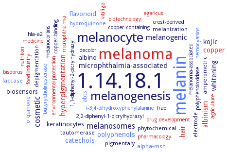
Word Map on EC 1.14.18.1 
-
1.14.18.1
-
melanin
-
melanoma
-
melanocyte
-
melanogenesis
-
melanosomes
-
cosmetic
-
microphthalmia-associated
-
hyperpigmentation
-
melanogenic
-
catechols
-
polyphenols
-
kojic
-
copper
-
albinism
-
whitening
-
hair
-
keratinocytes
-
albino
-
flavonoid
-
biosensors
-
o-quinone
-
amperometric
-
phytochemical
-
pigmentary
-
vitiligo
-
tautomerase
-
laccase
-
abts
-
alpha-msh
-
melanization
-
depigmentation
-
hydroquinone
-
electrode
-
melanocortins
-
2,2-diphenyl-1-picrylhydrazyl
-
copper-binding
-
polyphenoloxidase
-
frap
-
biotechnology
-
agriculture
-
pharmacology
-
hla-a2
-
nutrition
-
1,1-diphenyl-2-picrylhydrazyl
-
food industry
-
hemocyanins
-
environmental protection
-
l-3,4-dihydroxyphenylalanine
-
crest-derived
-
drug development
-
microphthalmia
-
medicine
-
agaricus
-
butyrylcholinesterase
-
melanoma-associated
-
copper-containing
-
decolor
-
bisporus
- 1.14.18.1
- melanin
- melanoma
- melanocyte
-
melanogenesis
- melanosomes
-
cosmetic
-
microphthalmia-associated
- hyperpigmentation
-
melanogenic
- catechols
- polyphenols
-
kojic
- copper
- albinism
-
whitening
- hair
-
keratinocytes
-
albino
- flavonoid
-
biosensors
- o-quinone
-
amperometric
-
phytochemical
-
pigmentary
- vitiligo
-
tautomerase
- laccase
- abts
- alpha-msh
-
melanization
-
depigmentation
- hydroquinone
-
electrode
-
melanocortins
-
2,2-diphenyl-1-picrylhydrazyl
-
copper-binding
- polyphenoloxidase
-
frap
- biotechnology
- agriculture
- pharmacology
-
hla-a2
- nutrition
-
1,1-diphenyl-2-picrylhydrazyl
- food industry
- hemocyanins
- environmental protection
- l-3,4-dihydroxyphenylalanine
-
crest-derived
- drug development
- microphthalmia
- medicine
- agaricus
- butyrylcholinesterase
-
melanoma-associated
-
copper-containing
-
decolor
- bisporus
Reaction
2 L-dopa
+
Synonyms
AbPPO1, AbPPO4, AbTYR, aurone synthase, catalase-phenol oxidase, catechol oxidase, catecholase, CATPO, chlorogenic acid oxidase, chlorogenic oxidase, cresolase, cresolase/monophenolase, CsPPO, CZA14Tyr, deoxy-tyrosinase, dihydroxy-L-phenylalanine:oxygen oxidoreductase, Diphenol oxidase, diphenolase, dopa oxidase, EC 1.10.3.1, EC 1.14.17.2, Hc-derived phenoloxidase, Hc-phenoloxidase, HcPO, HdPO, hemocyanin-derived phenoloxidase, jrPPO1, jrTYR, L-DOPA monophenolase, L-DOPA oxidase, L-DOPA:oxygen oxidoreductase, L-tyrosine hydroxylase, MdPPO1, melC2, MelC2 tyrosinase, met-tyrosinase, monophenol dihydroxyphenylalanine:oxygen oxidoreductase, monophenol monooxidase, monophenol monooxygenase, monophenol monoxygenase, monophenol oxidase, monophenol oxygen oxidoreductase, monophenol, 3,4-dihydroxy L-phenylalanine (L-DOPA):oxygen oxidoreductase, monophenol, dihydroxy-L-phenylalanine oxygen oxidoreductase, monophenol, dihydroxy-L-phenylalanine:oxygen oxidoreductase, monophenol, dihydroxyphenylalanine:oxygen oxidoreductase, monophenol, L-Dopa: oxidoreductase, monophenol, L-DOPA: oxygen oxidoreductase, monophenol, o-diphenol: oxygen oxidoreductase, monophenol, o-diphenol:O2 oxidoreductase, monophenol, o-diphenol:oxygen oxido-reductase, monophenol, o-diphenol:oxygen oxidoreductase, monophenol, polyphenol oxidase, monophenol: dioxygen oxidoreductases, hydroxylating, monophenolase, monphenol mono-oxygenase, More, mTyr, murine tyrosinase, mushroom tyrosinase, mushroom tyrosine, N-acetyl-6-hydroxytryptophan oxidase, o-diphenol oxidase, o-diphenol oxidoreductase, o-diphenol oxygen oxidoreductase, o-diphenol: O2 oxidoreductase, o-diphenol: oxidoreductase, o-diphenol:O2 oxidoreductase, o-diphenol:oxygen oxidoreductase, o-diphenolase, OCA1, Orf13, oxygen oxidoreductase, phenol oxidase, phenol oxidases, phenolase, phenoloxidase, PO, polyaromatic oxidase, polyphenol oxidase, polyphenol oxidase 3, polyphenol oxidase 4, polyphenol oxidase B, polyphenolase, polyphenoloxidase, PotPPO, PPO, PPO 3, PPO B, PPO1, PPO2, PPO3, pro-PO III, prophenoloxidase III, pyrocatechol oxidase, SPRTyr, ST94, ST94t, tryosinase, tryrosinase, TY, tyr, TYR1, TYR2, tyrA, TyrBm, tyrosinase, tyrosinase 2, tyrosinase 4, tyrosinase diphenolase, tyrosine-dopa oxidase


 results (
 results (
 top
top


