1.14.17.1: dopamine beta-monooxygenase
This is an abbreviated version!
For detailed information about dopamine beta-monooxygenase, go to the full flat file.

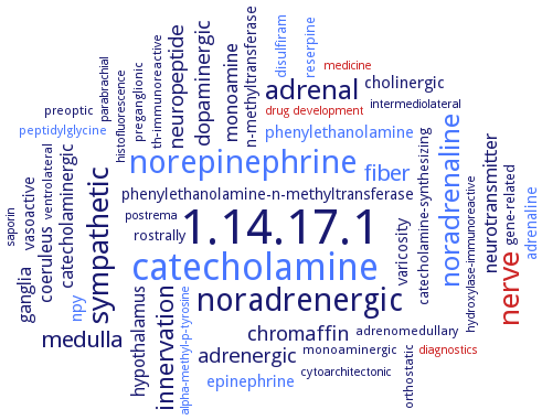
Word Map on EC 1.14.17.1 
-
1.14.17.1
-
catecholamine
-
noradrenergic
-
norepinephrine
-
nerve
-
sympathetic
-
noradrenaline
-
adrenal
-
innervation
-
medulla
-
fiber
-
dopaminergic
-
neuropeptide
-
chromaffin
-
adrenergic
-
monoamine
-
neurotransmitter
-
ganglia
-
coeruleus
-
hypothalamus
-
catecholaminergic
-
phenylethanolamine
-
npy
-
epinephrine
-
cholinergic
-
adrenaline
-
phenylethanolamine-n-methyltransferase
-
n-methyltransferase
-
varicosity
-
vasoactive
-
rostrally
-
reserpine
-
gene-related
-
catecholamine-synthesizing
-
disulfiram
-
peptidylglycine
-
preganglionic
-
adrenomedullary
-
th-immunoreactive
-
orthostatic
-
preoptic
-
ventrolateral
-
monoaminergic
-
parabrachial
-
saporin
-
intermediolateral
-
hydroxylase-immunoreactive
-
cytoarchitectonic
-
medicine
-
drug development
-
postrema
-
diagnostics
-
histofluorescence
-
alpha-methyl-p-tyrosine
- 1.14.17.1
- catecholamine
-
noradrenergic
- norepinephrine
- nerve
-
sympathetic
- noradrenaline
- adrenal
-
innervation
- medulla
- fiber
-
dopaminergic
-
neuropeptide
-
chromaffin
-
adrenergic
-
monoamine
-
neurotransmitter
- ganglia
- coeruleus
- hypothalamus
-
catecholaminergic
- phenylethanolamine
- npy
- epinephrine
-
cholinergic
- adrenaline
-
phenylethanolamine-n-methyltransferase
- n-methyltransferase
-
varicosity
-
vasoactive
-
rostrally
- reserpine
-
gene-related
-
catecholamine-synthesizing
- disulfiram
- peptidylglycine
-
preganglionic
-
adrenomedullary
-
th-immunoreactive
-
orthostatic
-
preoptic
-
ventrolateral
-
monoaminergic
-
parabrachial
- saporin
-
intermediolateral
-
hydroxylase-immunoreactive
-
cytoarchitectonic
- medicine
- drug development
- postrema
- diagnostics
-
histofluorescence
- alpha-methyl-p-tyrosine
Reaction
Synonyms
3,4-dihydroxy-phenylethylamine, ascorbate: oxygen oxidoreductase (3-hydroxylating), 3,4-dihydroxyphenethylamine beta-oxidase, 3,4-dihydroxyphenylethylamine beta-hydoxylase, 4-(2-aminoethyl)pyrocatechol beta-oxidase, DbetaH, DbetaM, DBH, DBM, Dopa beta-hydroxylase, dopamine beta hydroxylase, dopamine beta-hydrolase, dopamine beta-hydroxylase, dopamine beta-mono-oxygenase, dopamine beta-monooxygenase, dopamine beta-oxidase, dopamine hydroxylase, dopamine(3,4-dihydroxyphenethylamine)beta-mono-oxygenase, dopamine-B-hydroxylase, dopamine-beta hydroxylase, dopamine-beta-hydroxylase, dopamine-beta-monooxygenase, EC 1.14.2.1, gDBH, LvDBH, MDBH, oxygenase, dopamine beta-mono-, pDbetaH, phenylamine beta-hydroxylase, plasma DbetaH activity, plasma dopamine beta-hydroxylase, plDbetaH, SDBH, TBetaM, tyramine beta-monooxygenase


 results (
 results (
 top
top


