1.13.11.52: indoleamine 2,3-dioxygenase
This is an abbreviated version!
For detailed information about indoleamine 2,3-dioxygenase, go to the full flat file.

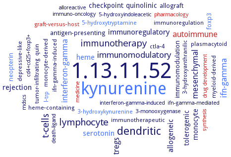
Word Map on EC 1.13.11.52 
-
1.13.11.52
-
kynurenine
-
dendritic
-
t-cells
-
lymphocyte
-
immunotherapy
-
ifn-gamma
-
immunomodulatory
-
autoimmune
-
mesenchymal
-
interferon-gamma
-
tregs
-
allogeneic
-
immunoregulatory
-
rejection
-
serotonin
-
heme
-
monocyte
-
tolerogenic
-
quinolinic
-
checkpoint
-
neopterin
-
allograft
-
immunomodulation
-
foxp3
-
antigen-presenting
-
immunoregulation
-
myeloid-derived
-
3-hydroxykynurenine
-
ctla-4
-
graft-versus-host
-
l-trp
-
3-hydroxyanthranilic
-
quin
-
tumor-infiltrating
-
heme-containing
-
depressive-like
-
immunotherapeutic
-
plasmacytoid
-
5-hydroxytryptamine
-
monocyte-derived
-
ifn-gamma-induced
-
5-hydroxyindoleacetic
-
interferon-gamma-induced
-
immuno-oncology
-
drug development
-
cd4+cd25+foxp3+
-
mdscs
-
pharmacology
-
alloreactive
-
3-monooxygenase
-
ifn-gamma-mediated
-
medicine
-
death-ligand
-
synthesis
- 1.13.11.52
- kynurenine
- dendritic
- t-cells
- lymphocyte
-
immunotherapy
- ifn-gamma
-
immunomodulatory
- autoimmune
- mesenchymal
- interferon-gamma
-
tregs
-
allogeneic
-
immunoregulatory
-
rejection
- serotonin
- heme
- monocyte
-
tolerogenic
-
quinolinic
-
checkpoint
- neopterin
-
allograft
-
immunomodulation
- foxp3
-
antigen-presenting
-
immunoregulation
-
myeloid-derived
- 3-hydroxykynurenine
-
ctla-4
- graft-versus-host
- l-trp
-
3-hydroxyanthranilic
-
quin
-
tumor-infiltrating
-
heme-containing
-
depressive-like
-
immunotherapeutic
-
plasmacytoid
- 5-hydroxytryptamine
-
monocyte-derived
-
ifn-gamma-induced
-
5-hydroxyindoleacetic
-
interferon-gamma-induced
-
immuno-oncology
- drug development
-
cd4+cd25+foxp3+
-
mdscs
- pharmacology
-
alloreactive
-
3-monooxygenase
-
ifn-gamma-mediated
- medicine
-
death-ligand
- synthesis
Reaction
Synonyms
31854, BRAFLDRAFT_126354, CG5163, EC 1.13.1.12, hIDO, hIDO1, hTDO, IDO, IDO-1, IDO-2, IDO-I, IDO-II, IDO-III, IDO-IV, IDO1, IDO2, INDO, INDOL1, indolamine 2,3-dioxygenase, indole 2,3-dioxygenase, indoleamine 2, 3-dioxygenase, indoleamine 2,3 dioxygenase, indoleamine 2,3-dioxygenase, indoleamine 2,3-dioxygenase 1, indoleamine 2,3-dioxygenase 2, indoleamine 2,3-dioxygenase-1, indoleamine 2,3-dioxygenase-2, indoleamine 2,3-dioxygenase-like protein, indoleamine-2,3-dioxygenase, Indoleamine-pyrrole 2,3-dioxygenase, L-tryptophan 2,3-dioxygenase, L-tryptophan pyrrolase, mIDO, oxygenase, tryptophan 2,3-di-, proto-IDO, proto-indoleamine 2,3-dioxygenase, superoxygenase, TDO, TDO2, TioF, TO, TRPO, Tryptamin 2,3-dioxygenase, tryptamine 2,3-dioxygenase, tryptophan 2,3-dioxygenase, tryptophan dioxygenase, tryptophan oxygenase, tryptophan peroxidase, tryptophan pyrrolase, tryptophan-2,3-dioxygenase, tryptophanase, v1g244579, Vermilion protein


 results (
 results (
 top
top


